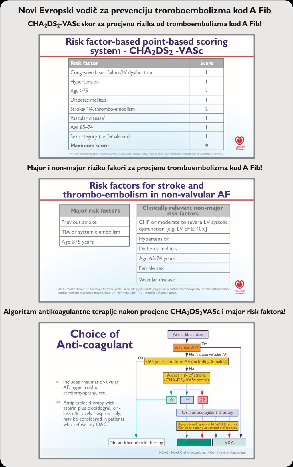
Content Medical Journal (2016) Vol. 22, No 3 Original article Smoking rate among health care professionals employed in the University Clinical Center
|
|
|
- Василије Михаиловић
- пре 5 година
- Прикази:
Транскрипт

1

2

3
4


5
6
7
8
9
10
11
12 Content Medical Journal (2016) Vol. 22, No 3 Original article Smoking rate among health care professionals employed in the University Clinical Center Sarajevo 115 Sebija Izetbegović, Amer Ovčina EUTOS score as predictor of event free survival in patients with CML Ph+ in early and late chronic phase in TKI era 123 Alma Sofo-Hafizović, Emina Fazlibegović, Refet Gojak, Lejla Ibričević-Balić Is pertussis a forgotten disease? Pertussis in infants admitted to the Pediatric Clinic of the University Clinical Center Sarajevo 130 Selma Dizdar, Belma Paralija, Edo Hasanbegović, Ganimeta Bakalović, Amra Džinović, Verica Mišanović, Jasmina Fočo-Solak Evaluation of myocardial perfusion defects in patients with anatomical assessment of coronary stenosis 134 Aida Hasanović Beta 2 microglobulin as prognostic factor in newly diagnosed myeloma patients 137 Lejla Burazerović, Edo Hasanbegović Professional article Subcutaneous central venous port implantation under fluoroscopy and ultrasound guidance 140 Vesna Đurović-Sarajlić, Elma Kapisazović, Jasmina Redžepagić, Sanela Vesnić, Aladin Čarovac, Nihad Kukavica Review article Prof. dr. Aleksandar Terzin the first director of the Institute of Virology and Immunology of the Faculty of Medicine, University of Sarajevo ( ) 146 Šukrija Zvizdić Case report Combined use of psychopharmacotherapy and cognitive behavioral psychotherapeutic techniques in the treatment of social phobia of a patient diagnosed with social anxiety disorder 149 Alem Ćesir, Amir Balić Instructions to authors 151 Uputstva autorima 153
13 Medical Journal (2016) Vol. 22, No 3, Original article Smoking rate among health care professionals employed in the University Clinical Center Sarajevo Učestalost pušenja duhana kod radnika u zdravstvu Sebija Izetbegović 1*, Amer Ovčina 2 1University Clinical Center Management, Bolnička 25, Sarajevo, Bosnia and Herzegovina 2Discipline for Clinical Science and Education, Office of Clinical Quality Improvement and Healthcare Safety, Bolnička 25, Sarajevo, Bosnia and Herzegovina *Corresponding author ABSTRACT Tobacco smoking is the single most significant risk factor for health in the European region. According to the World Health Organization reports the number of smokers in the world has increased and it is expected that by the number of smokers in the world will increase from current 1.3 to 1.7 billion. It is estimated that 50% of adult population of Bosnia and Herzegovina are smokers. Smoking tobacco among health care profesionals is a public health and ethical problem and it is a risk factor in the development of a large number of non-communicable diseases of respiratory, cardiovascular, digestive, reproductive, nervous and other systems. Tobacco smoking causes three types of addictions, namely physical, psychological and metabolic, and therefore according to the International Classification tobacco smoking has been included among mental and behavioural disorders. Study objective: to evaluate smoking rate among health care workers, to examine and analyze the smoking habits of health care workers, and to demonstrate correlation with previously conducted studies. Materials and methods: this descriptive data analysis study included 378 respondents/employees of the Clinical Centre University of Sarajevo. A questionnaire related to smoking status, taken from the European Network of Smoke-Free Hospitals (Indicators for Hospitals, AKAZ, 2014) was used as an instrument of this descriptive study. The questionnaire was anonymous. The study was conducted in the period from 1 October to 30 November Results: the study showed that out of the total of 378 respondents 129 (34%) respondents consumed tobacco products on a daily basis, and 46 (12%) respondents only occasionally. Majority of smokers, that is 103 (64%), consumed tobacco products during working hours. Total of 101 (63%) respondents expressed their wish to give up smoking and 355 (93%) of them considered that the CCUS did not have services which could contribute to their wish to give up smoking. Discussion and conclusion: the continuous research related to the prevalence of smoking habits conducted among the health care workers of the Clinical Centre University of Sarajevo in the period from 2014 to 2016 confirmed that out of the total number of respondents the number of regular smokers ranged from 40% to 50%. Factors influencing the prevalence of tobacco use in the population of Bosnia and Herzegovina include: easy access to tobacco products, lack of restrictions on the sale of tobacco products to minors, wide sale network and the lack of inspections. In order to achieve the Goals for Health in the 21st century set by the World Health SAŽETAK Duhan je najvažniji pojedinačni faktor rizika za zdravlje u regiji Evrope. Prema podacima Svjetske zdravstvene organizacije broj pušača u svijetu se povećava tako da se procjenjuje da će do Godine sa današnjih 1,3 milijarde, broj ovisnika o cigareti porasti na 1,7 milijardi. Procjenjuje se da u BiH duhan konzumira 50% odraslog stanovništva. Pušenje duhana kod zdravstvenih radnika predstavlja javno zdravstveni i etički problem. Pušenje duhana je faktor rizika u nastanku velikog broja nezaraznih oboljenja respratornog, kardiovaskularnog, digestivnog, reproduktivnog, nervnog i drugih sistema. Uživanje duhana izaziva tri tipa ovisnosti i to fizičku, psihičku i metaboličku, te je zbog toga međunarodnom klasifikacijom bolesti pušenje duhana uvršteno među mentalne poremećaje i poremećaje ponašanja. Cilj rada: utvrditi stopu pušenja duhana kod radnika u zdravstvu; ispitati i analizirati pušačke navike kod radnika u zdravstvu; prikazati korelativnu vezu sa ranije provedenim istraživanjima. Materijali i metode: istraživanjem je obuhvaćeno 378 ispitanika/radnika zaposlenih u Kliničkom centru Univerziteta u Sarajevu. Istraživanje je deskriptivna analiza podataka. Kao instrument za deskriptivno istraživanje korišten je anketni upitnik o pušačkom statusu, preuzet od strane Evropske mreže bez duhanskog dima u bolnicama (Indikatori za bolnice, AKAZ, 2014). Anketni upitnik je bio anoniman i iz upitnika se ne može saznati identitet ispitanika. Istraživanje je provedeno u periodu od do godine. Rezultati istraživanja: istraživanje je pokazalo je da od ukupno 378 ispitanika, 129 (34%) ispitanika konzumira duhanske proizvode svakodnevno, a 46 (12%) povremeno. Među ispitanicima pušačima je bio najveći broj ispitanika koji duhanske proizvode konzumira za vrijeme rada i to 103 (64%) ispitanika. Želju za prestankom pušenja duhana izrazio je 101 (63%) ispitanik, ali njih 355 (93%) smatraju da KCUS nema servis koji će im pomoći u odvikavanju od loše navike. Diskusija i zaključak: kontinuiranim istraživanjem raširenosti pušačkih navika u KCUS u periodu od do godine potvrđeno je da se broj stalnih pušača kreće od 40% do 50% u ispitivanim grupama. Faktori koji utiču na raširenost upotrebe duhana kod populacije u BiH su lahka dostupnost duhanskim prerađevinama, nepostojanje ograničenja prodaje duhanskih prerađevina maloljetnim licima, široka mreža prodaje i nedostatak inspekcijskih kontrola. U cilju ispunjenja Ciljeva zdravlja za sve u 21. stoljeću koje je propisala Svjetska zdravstvena organizacija neophodno je kontinuirano raditi na smanjenju stope pušenja duhana među radnicima u zdravstvu.
14 S. Izetbegović et al. Organization it is necessary to continue efforts in reducing the rate of smoking among health care workers. Ključne riječi: duhan, pušenje, navike, učestalost, prevencija, radnici u zdravstvu Key words: tobacco, smoking, habits, incidence, prevention, health care workers INTRODUCTION Tobacco smoking is a common non-infectious and social disease which damages the physical, mental health and socially well-being, which is in fact objective physical, psychological and biochemical ascertainment (1). World Health Organization classifies tobacco smoking among mental and behavioral disorders within International Classification of Diseases, and marked it with the code F 17. Health authorities consider the enjoyment of tobacco as unhealthy habit and maladaptation (2). Despite scientific evidence that tobacco smoking is the single most significant risk factor for health in the Region of Europe, smoking habits remain high, especially among the younger population. Smoking rates differ by region and country and also depends on the economic situation of the nation (3). According to the World Health Organization, smoker is addicted to cigarettes, a person smokes per day more than twenty cigarettes. Cigarettes shortens life on average for ten to twelve years. Smokers use health care for the treatment of diseases caused by smoking (4). The result show that prevalence of tobacco consumption is in underdeveloped and developing countries. As tobacco use has declined in high-income, more-developed countries, it is now escalating in lessdeveloped countries. Each year, tobacco causes 3.5 million deaths worldwide and it is expected to grow up to 10 million annually by 2020, with 7 million deaths in developed countries. Fifty
15 percent of regular smokers will die from cigarettes in their middle age (5). World Health Organization experts consider that more than 25 are smoking-related diseases. The same researchers say giving up smoking before 35 years of age will increase a chance for exsmokers to live as long as those who have never enjoyed tobacco (6).Tobacco smoke increases the risk of many cancers, chronic heart disease, low birth weight, sudden infant death, allergies and other health problems (7). EU/EFTA analysis shows that tobacco products cost the EU countries at least 100 billion Euros a year, causing unprecedented suffering for smokers, their families and friends; represent an enormous cost to the economy; and are responsible for environmental destruction and fires. Significant economic losses due to tobacco use are primarily related to the early completion of productive life and high costs of treatment (8). Legislation in Federation of Bosnia and Herzegovina is solid basis for the controlled supervision of production, transport and consumption of tobacco products and are in compliance with international recommendations and documents. The FBiH legislation currently in force includes: Act on Limited use of Tobacco Products (FBiH Official Gazette no. 6/95, 6 / 98, 35/98, 11/99, 13/00, 52/01), Excise Duties on Tobacco Products, the Regulations on Printing, Payment, Recording and Handling of Tax Stamps for Marking Tobacco Products, the FBiH Law on Tax Administration, Guidelines for Realizing Financial Incentives in the Primary Agricultural Production and the Tobacco Act. Most of the legal norms concerning the control, production and sale of tobacco products are contained in the Act on Limited Consumption of Tobacco Products. Difficulties arise in the implementation of the law provisions, due to absence of clear mechanisms for control inspection (1,6). In accordance with the recommendations of the Framework Convention on Tobacco Control by WHO, Federal Ministry of Health initiated activities in 2010 that health care workers must be committed to the prevention, early detection and control of risk factors to human health, and also harmonization of legislation through the adoption of the Law on Health Care of the Federation of Bosnia and Herzegovina. The Law on the rights, obligations and responsibilities of patients introduces the right of patients and needs to be educated about their bad habits and personal responsibility of for their own health in terms of practicing healthy habits and the axiom of choice (8,9). Considering commitment of Bosnia and Herzegovina, taken over by ratifying the Framework Convention for Tobacco Control of the World Health Organization it launched an initiative to control tobacco, which main objective is in the new Law on Tobacco Control, which is harmonized with the Framework Convention of the World Health Organization and the European Union directives. The law was supported by the relevant federal ministries, Health Insurance Institute, Campaign for Tobacco Free Kids and non-governmental organizations dealing with the problem of tobacco smoking in Bosnia and Herzegovina. The new Law is in the process of adoption and enactment into force (10). Reducing the smoking rate is a complex social issue that requires binding wide access. Health care workers, and especially health professionals (doctors and nurses-technicians) have a great influence on the attitudes and behavior of patients and generally in the long term and the overall health of the nation. Smoking habits of employees in health institutions is a mirror of the situation, the impact and success of the general policy of tobacco control in the country (5,11). Research objectives 1. To establish the smoking rate among health care workers 2. To examine and analyze the smoking habits in health care workers 3. To show correlation with previously conducted studies MATERIALS AND METHODS The study included 378 respondents or 10% of employees in all 45 organizational units of the University Clinical Center Sarajevo. Simple random sampling provided equal representation of respondents by gender, education and age. The descriptive research used questionnaire related to smoking status, taken by the European Network of Smoke-Free Hospitals (Indicators for Hospitals, AKAZ 2014).The questionnaire was anonymous.the questionnaire was created in the software program Toluna QuickSurveys and through link connection available to all employees in organizational units. The survey was conducted in the period from 1 October to 30 November All data were processed in Microsoft Excel and results are displayed in tables and figures. Figure 1 Age of respondents.


16 S. Izetbegović et al. Figure 2 Gender. Figure 5 Smoking status in Figure 3 Level of education. Figure 6 Smoking status in Figure 4 Work position. Figure 7 Smoking status in 2016.


17 Smoking rate among health care professionals employed in the University Clinical Center Sarajevo Figure 8 How soon after you wake up do you smoke your first cigarette? Figure 11 How many times did you try to give up smoking. Figure 9 When did you first start smoking? Figure 12 Reasons to quit smoking. Figure 10 Why did you start smoking? Figure 13 Number of daily smoked cigarettes.

18 S. Izetbegović et al. Figure 14 Do you think that the Clinical Center could help its employees to quit smoking? Figure 17 Do you plan to quit smoking and when? Figure 15 How many times did you try to quit smoking? Figure 18 Are your parents smokers? Figure 16 Reasons for failure to give up smoking. Figure 19 Are your colleagues smokers?


19 Smoking rate among health care professionals employed in the University Clinical Center Sarajevo Figure 20 Do you know that smoking causes serious consequences to your health? Figure 23 Do you work at night? Figure 21 Do you have any health problems caused by smoking? Figure 24 Do you smoke at workplace? Figure 22 Did your colleagues advice you to quit smoking? Figure 25 Are there rooms provided for smoking at your workplace?
20 Figure 26 Is there a Quit Smoking Treatment Service at the CCUS? DISCUSSION The study on smoking habits among health care workers of the University Clinical Center Sarajevo has been conducted since 2014 in the context of the implementation of the system of quality and safety of health services. In 2014, the study on the smoking rate among health care professionals was conducted on the sample of 563 (15.1%) employees, of which 217 were males and 346 females. The study showed that 41% of respondents were smokers. In 2015, the survey was conducted on 479 respondents, and the results shown that 43% of respondents were smokers. Studies have shown that mostly smokers are health care professionals employed at stressful positions, such as surgery and emergency block of the CCUS. In 2016, the survey was conducted on 378 respondents and the study showed that 129 (34%) respondents were regular daily smokers and that 46 (12%) smoke tobacco products occasionally. Majority of smokers (64%) consume tobacco products during working hours. Smoking tobacco is prohibited in the hospital premises which causes anxiety among workers. The Accreditation Standards for Hospitals require Smoke-Free Hospital. Given that the CCUS is an institution in the accreditation process it is required to consistently implement the standards. Smoking area is provided within the building on the site specified for smokers. This is one of the main reasons for health care workers to quit smoking. Fortunately, 139 (86.88%) respondents stated that they did not experience health problems caused by smoking tobacco products, and results are similar to researches conducted in previous years. According to the 2002 and 2003 surveys conducted in the Department of Public Health on a sample of 3020 respondents, 37.6% of them were daily smokers (49.2% men and 29.8% women). Smoking habits and increase of malignant (cancerous) diseases in the Federation are also on the rise. For example, in 1998 there were 710 deaths, in 1999 that number increased to 714; in 2000 there were 833 deaths, and in 2001 that number increased to 846. In Bosnia and Herzegovina, smokers monthly spend between BAM on cigarettes, which annually amount to about 1800 BAM. It is estimated that Bosnians spend over a billion Euros on cigarettes annually (6). According to the latest research on health condition of adult population in the Federation in 2012, conducted by the Institute of Public Health of the Federation of Bosnia and Herzegovina, it was shown that tobacco smoking was the leading addiction disease in the Federation of Bosnia and Herzegovina. The study showed that there was 41.1% of permanent smokers in the Federation of Bosnia and Herzegovina, of which 56.3% were men and 31.6% women. Smoking among young people is also particularly widespread and research showed that 12.7% of young people aged years consumed tobacco products (12). S. Izetbegović et al. In the Republic of Croatia, the number of smokers is 1.6 million, of which in Zagreb. From the perspective of gender, results showed that 34% of men and 36% women regularly smoke tobacco. Each year in Croatia dies between 12,000 to 15,000 people as a consequence of smoking (13). A survey conducted in 2011 in the Koprivnica-Križevci County, showed that the prevalence of smokers among all employees in health institutions was 26.4%, equal for both medical and nonmedical staff. The prevalence of smokers was lowest among physicians (19.4%) and the highest among nurses (29.4%), especially nurses with secondary level of education (30.4%). More smokers was found among women and people younger than 45 years than among men and people older than 45 years (14). According to the number of smokers, Serbia is highly ranked in the European and world scale. According to the WHO, 47% of Serbia population are smokers, and according to a research conducted in 2006 it was 33.6%. A survey conducted among health care workers in health institutions of Serbia in 2006 showed the following: out of the total of respondents, 45.6% were smokers. Minority of them were physicians (34.13%), and majority related to nurses (51.87%). This data indicates that majority of smokers among the respondents related to nurses. Tobacco products consumed 46.4% of male and 45.4% female respondents. There were significant differences in attitude about the role of health professionals in smoking cessation between both smokers and non-smokers, and between doctors and nurses (15). Our study was conducted in 2014, 2015 and 2016 and it also showed that majority of smokers related to nurses, especially those working in night shifts (30%). Quitting smoking brings to the individual physical, psychological and economic well-being. This is an important step towards reducing the health risk they are exposed to. The most common motives for quitting smoking are economic factors, aesthetic and anti-advertising (16,17). Quitting smoking is a cyclic process comprising the following steps: thinking on termination, the decision on termination, attempting to quit and maintain the status of former smokers. Less than 5% of smokers directly cross the confirmed status of former smokers without recurrence. Factors that encourage individuals to quit smoking are: reduced social acceptability of smoking, increased health care, increased price of tobacco products and so on. The factors that encourage recurrence of smoking are: desire for nicotine, weight gain, social pressures etc. (16). As a reason for failure to quit smoking, respondents (21 of them or 51%) mostly cited the problem of smokers around the work environment.
21 Smoking rate among health care professionals employed in the University Clinical Center Sarajevo The positive side of our research is that respondents, smokers
22 express a desire to quit smoking (101 of them or 63%), and 355 of them (93%) believe that the CCUS does not have service that will help them quit smoking. CONCLUSION Although it is proven that consumption of tobacco affects human health, it is more widespread among health care professionals, especially among health care workers as shown in numerous researches conducted in Bosnia and Herzegovina. Research on smoking rate conducted at the CCUS in the period showed that the number of regular smokers among respondents ranged from 40% to 50%. Factors affecting the spreading of smoking among the population of Bosnia and Herzegovina are easy access to tobacco products, lack of restrictions on the sale of tobacco products to minors, a wide network of sales and lack of inspections. Studies conducted at the CCUS in 2014, 2015 and 2016 showed that there was a prevalence of smokers among workers of younger population, predominantly female population and workers performing stressful jobs. Health care workers are ethically and morally accountable primarily to their own health, and then for the health of their clients, so they should be advocates of mass campaigns against smoking. The hospital is a place where smoking is prohibited, and where rights of patients and non-smokers among employees must be appreciated. In order to achieve the Goals for Health in the 21st century, it is essential that health care workers be actively involved in abandoning the bad habits of smoking and in promoting healthy lifestyles. S. Izetbegović et al. 12. Kulčar Ž, Kovačić L, Bedenić B. Rasprostranjenost pušenja u stanovništvu SR Hrvatske. Liječ. Vjesn. 1974;96: Gazdek D. Trend navike pušenja u zdravstvenim ustanovma Koprivničko križevačke županije i politika kontrole pušenja. Komparativna studija g., Zavod za javno zdravstvo Koprivničko-križevačke županije, Koprivnica, Stojanović M, Mušinović D, Petrović B, Milošević Z, Milosavljević I, Višnjić A, et al. Pušačke navike, znanje i stavovi o pušenju zaposlenih u zdravstvenim institucijama u Srbiji. Vojnosanitetski pregled. 2013;70(5): Ovčina A. Savremene metode odvikavanja od pušenja duhana. Bilten «Trezvenost», Vol. 7/8. Sarajevo, Omanić A, Omanić J, Pećanac N. Vodič za odvikavanje od pušenja duhana. Sarajevo, Reprint requests and correspondence: Sebija Izetbegović, MD, PhD General Manager University Clinical Center Sarajevo Bolnička 25, Sarajevo Bosnia and Herzegovina kagd@kcus.ba Conflict of interest: none declared. REFERENCES 1. Omanić A. Javno-zdravstveni problemi pušenja duhana i njihova prevencija. U: Priručnik za multidisciplinarni pristup prevenciji zloupotrebe psihoaktivnih supstanci. Udruženje za prevenciju ovisnosti i smanjenje štete, Sarajevo, Link Zdravlje 21. Zdravlje za sve u 21 stoljeću. Zavod za zdravstvenu zaštitu BiH, Sarajevo World Health Organization (WHO). WHO Report on the Global Tobacco Epidemic, Warning about the dangers of tobacco. Geneva: WHO, Ovčina A, et al. Bioetički pristup problemu pušenja duhana u Bosni i Hercegovini. U: Zbornik radova 5. Lošinjski dani bioetike. Mali Lošinj, juna Strategija kontrole duhana u Federaciji BIH. Federalno ministarstvo zdravstva, Zavod za javno zdravstvo FBIH. Sarajevo Dilić M, Raljević E, et al. Trendovi kardiovaskularnih bolesti u BiH i nekim evropskim zemljama. Med Arh. 2004;58 (Suppl 1): Ramić-Ćatak A. Pušenje kao vodeća bolest ovisnosti na globalnom planu. posjećeno Zdravstveno statistički godišnjak Zavoda za javno zdravstvo FBiH, 2014.g. Zavod za javno zdravstvo FBiH.Godina XIII, Broj XIII. 9. Izvještaj o aktivnostima u KCUS. KCUS podržava unaprijeđenje legislative za kontrolu duhana u FBiH, Petak 01. jul/srp Omanić A, Nikšić D, Kurspahić-Mujčić A, Džubur A. Medicinski fakultet u Sarajevu. Med Arh. 2004;58 (2 Suppl 1): Zdravstveno stanje stanovništva i zdravstvena zaštita u Federaciji Bosne i Hercegovine Zavod za javno zdravstvo FBiH, Sarajevo 2014.
23 Medical Journal (2016) Vol. 22, No 3, Original article EUTOS score as predictor of event free survival in patients with CML Ph+ in early and late chronic phase in TKI era EUTOS skor kao prediktor preživljavanja bez događaja kod pacijenata sa hroničnom mijeloičnom leukemijom Ph+ u ranoj i kasnoj hroničnoj fazi u TKI eri Alma Sofo-Hafizović 1*, Emina Fazlibegović 2, Refet Gojak 3, Lejla Ibričević- Balić 1 1Hematology Clinic, University Clinical Center Sarajevo, Bolnička 25, Sarajevo, Bosnia and Herzegovina 2Faculty of Medicine, University of Sarajevo, Čekaluša 90, Sarajevo, Bosnia and Herzegovina 3Clinic of Infectious Diseases, University Clinical Center Sarajevo, Bolnička 25, Sarajevo, Bosnia and Herzegovina *Corresponding author ABSTRACT Introduction: European Treatment and Outcomes Study (EUTOS) score divides patients with chronic myeloid leukemia (CML) into low and high risk groups. Aim: to reevaluate predictor significance of EUTOS score in relation to achieving event free survival (EFS): acceleration/blast transformation or death, major molecular response (MMR) and 10-year overall survival of CML Ph+. Material and methods: this was a retrospective study, following CML Ph+ patients in the period from 2001 to The study involved 48 patients aged 18 to 60 years, mean age 44.6 years, of which 26 (54%) males and 22 females (46%). There were 24 patients in a low risk group and 24 in a high risk group, who were treated with Tyrosine kinase inhibitors (TKI) in the first, second and third line of therapy. Results: it was confirmed that MMR response at 3 to 6 months depends of EUTOS score (low vs. high) 64.3% vs 20% p=0.014; and at 18 months 66.7% vs 20%, p= Median survival in the low score group was months, specifically 95% CI ( ), which was longer than in the high score group in which median survival was months or 95% CI ( ). Survival rate differences without EFS in the low and high risk groups of EUTOS score were dependent χ 2 =4.463, p= Difference in survival according to EUTOS score was not statistically significant χ 2 =2.49, p= Conclusion: EUTOS score was not predictive for outcome in 10 years overall survival, but had predictive achieving EFS, value in MMR at 3 to 6 months and at 18 months. Statistic analysis: assessment of the significance of differences (X 2 tests), Log Rank (Mantel-Cox) test, Kaplan-Meier curve of survival. Key words: CML, EUTOS score, EFS SAŽETAK Uvod: EUTOS skor odvaja pacijente sa hroničnom mijeloičnom leukemiom (HML) u grupu niskog i visokog rizika. Cilj: provjeriti prognostički značaj EUTOS scora u odnosu na preživljavanje bez događaja (faza akceleracije, blastna transformacija i smrt), veliki molekularni odgovor i desetogodišnje ukupno preživljavanje oboljelih od hronične mijeloične leukemije Ph+. Materijali i metode: istraživanje je retrospektivna analiza praćenja pacijenata sa HML Ph+ u periodu od do godina. Analizirano je 48 pacijenata starosne dobi od 18 do 60 godina, prosječne starosne dobi 44,6 godina, od kojih 26 (54%) muškaraca i 22 (46%) žene. Rezultati: potvrđeno je da veliki molekularni odgovor na 3 do 6 mjeseci zavisi od EUTOS skora (niski naspram visokog) 64,3%:2055, p=0,014, i na 18 mjeseci 66,7%:20%, p=0,013. Mediana preživljavanja u grupi niskog rizika iznosila je 134,5 mjeseca odnosno 95% CI (110,5-158,6 mjeseci), što je duže od grupe visokog rizika sa 102,7 mjeseci odnosno 95% CI (79,4-126 mjeseci). Razlika u vremenu preživljavanja prema EUTOS scoru u toku 10 godina nije statistički značajna, c2=2,49; p=0,114. Razlike u preživljavanju bez događaja između grupe niskog i visokog rizika EUTOS skora su zavisne, c2=4,463 p=0,035. Zaključak: EUTOS skor nije prediktor 10- ogodišnjeg preživjavanja, ali je prediktor preživljavanja bez događaja (faza akceleracije, blastne transformacije i smrti) i nivoa velikog molekularnog odgovora na 3 do 6 mjeseci i 18 mjeseci. Statistička analiza: procjena značajnosti razlika (X2 test), Long Rank (Mantel- Cox) test, Kaplan-Meier krivulja preživljavanja. Ključne riječi: HML, EUTOS scor, EFS
24 A. Sofo-Hafizović et al. INTRODUCTION Chronic myeloid leukemia (CML) is hematopoietic stem cell clonal disease characterized by massive myeloid expansion. CML occurs most frequently in middle and older age, but sometimes it appears in younger population as well as in children. It represents around 20% of all leukemia with incidence of 1 in , with male predominance. Incidence varies for 0.6 to 2.0 cases in per year, increasing with age. Geographical and/or ethnical differences may contribute to various incidences within registries (1).
25 EUTOS score as predictor of event free survival in patients with CML Ph+ in early and late chronic phase in TKI era Until 30 years ago CML was a fatal disease with survival of
26 majority of patients up to 3 years. With revolutionary discovery of first and second generation of tyrosine kinas inhibitors (TKI) CML has been brought under control. Recent studies provide possibility of ceasing the therapy after certain period of time, when some patients are considered to be cured, but more studies are under way to verify this. European Leukemia Net and Novartis Oncology Europe have developed The European Treatment and Outcome Study (EUTOS) score as prognostic model to predict course of CML. Patients are divided into two groups, low risk and high risk group, based on prediction of progression of chronic phase into acceleration phase and blast crisis which in the end leads to lethal outcome. This prognostic model uses spleen size in centimeters and percentage of basophiles in peripheral blood (%) to sort patients into low risk (EUTOS score 87) or high risk group (EUTOS score > 87). A study that was basis for EUTOS score development had 2060 CML patient treated with I generation TKI, Imatinib. EUTOS score equation is (7 x basophile [%]) + (4 x spleen [cm]) (2). Providing the most effective treatment for CML patients requires recognition of patients with poor prognosis which can be done using EUTOS score. This group of patients needs close monitoring and prompt change of treatment when required, all this ensuring maximal response. EUTOS score is very simple tool, because it uses only two parameters, spleen size and basofill count in peripheral blood, and it can be used in primary care clinic and is widely accessible in everyday practice. Every prognostic classification system should be able to identify patients with poor prognosis irrespective of whether they are a part of trial or not. Sokal score has been in use for almost 30 years and it has been confirmed in different clinical trials and studies, while EUTOS score is still beeing reexamined and requires further studies that are under way. Achieving a cytogenetic answer to the therapy in the first performed studies was accepted as the most significant criteria for treatment evaluation in patients with CML as it is connected with improved survival and decsreased risk of disease transformation into the acceleration phase or blast crisis. Studies have also shown that the depth of achieved answer is not of sole importance but the time in which it is achieved as well. Quantitative RQ-PCR for examining BCR-ABL transcript is the most sensitive way for evaluation and minimal detection of BCR-ABL transcription in patients with CML for predictiong future results (3). In their study Yoshinori Shinohara et al. have concluded that the early MMR is an important predictor for forecasting long-term result of the imatinib treatment of patients with CML. Patients with BCR/ABL transcripts >10% for 6 months and >1% for 12 months had weaker EFS and a higher rate of desease progression into acceleration/blast transformation. On the other hand, patients who achieved MMR in 3-6 months had excellent responses, without progression into acceleration/blast transformation or death. The results of the research call attention to importance of achieving fast (early) MMR, as faster MMR has predictive impact on possibility of achieving further complete molecular response (CMR). CMR clearly comfirms the best outcome so it should in fact become the main goal of the therapy (4,5). MATERIALS AND METHODS Study population A. Sofo-Hafizović et al. Out of 87 patients diagnosed with CML 48 were included in the study, both male and female (male 26, female 22) age from 18 to 66. Patients included in the study were hospitalized, treated and followed up at the Hematology Clinic of the University Clinical Center Sarajevo in the period from 1 January 2001 to 31 December CML was diagnosed based on anamnesis, symptoms, fiscal status sings of disease, laboratory results of complete and differential blood count, cytomorphological analysis of bone marrow, FISH for bcr/abl transcript in bone marrow. Patients were treated with tyrosine kinas inhibitors, first and second generation in the first, second and third line of therapy. Patients not treated with TKI were excluded from the study. The study included 48 patients divided into 2 groups: EUTOS score low risk: 24 pts or 50% patients of both sexes (14 females and 10 males). EUTOS score high risk: 24 pts or 50% patients of both sexes (8 females and 16 males). This was a retrospective, clinical, comparative-descriptive study. Data was collected from the following medical charts: history of disease, clinical status, sex, age, spleen size - below left costal arch, and laboratory finding: basophile count - percentage in peripheral blood. Using the given data, spleen size and basophile count, EUTOS score was calculated and patients were divided into 2 groups (low risk and high risk), to find out whether there was a different pattern in disease progression (acceleration phase, blast crisis and death) and in 10-year survival. EUTOS score uses formula, 7 x basophile percentage (%)+ 4 x spleen size (cm), to set patients into one of the two groups (low risk EUTOS score 87 and high risk EUTOS score > 87). Total number of patients treated with the first generation of TKI Imatinib was 27 in the first and second line of a specific therapy, whereas 21 patients were treated with Nilotinib, the second generation of TKI in the first, second and third line of the specific therapy. Patients were evaluated in order to assess hematological response based on full blood count and spleen size every 15 days until they reached hematological remission; complete cytogenetic response was assessed in bone marrow determining number of copies of Ph+ metaphases using fluorescent situ hybridization (FISH) at 3, 6, 12 and more months and major molecular response was assessed using real time polymerase chain reaction (RT-Q- PCR) detecting BCRABL transcript in and 1 million cells from peripheral blood at 3, 6, 18 and more months. Criteria set by European Leukemia Net (ELN) group were used to define hematological, cytogenetic and molecular response, and criteria for acceleration phase of blast transformation. Disease course was followed regarding unwanted events: acceleration phase, blast crisis and death, in both low and high risk groups as well as in 10 years survival.


27 EUTOS score as predictor of event free survival in patients with CML Ph+ in early and late chronic phase in TKI era Statistical analysis Spleen size in the low risk group was 14+/-1.97 and 18,7+/-3,8 cm in the high risk group. The difference in the size was statistically Nominal and ordinal variables were analyzed using X 2 test. significant, p<0,0005. Survival and differences in survival between the groups was Table 1 Values Le, Ne, Ly, Mo, Eo, Ba and EUTOS score calculated using Log Rank (Mantel-Cox) c 2 test. Results were (low/high). displayed in Kaplan-Meier curve and survival tables. α=0.05 was statistical significance. During the research all ethical principles were followed in line with the Helsinki Declaration. RESULTS The study included 48 patients with CML Ph+in chronic phase who were treated with TKI at the Hematology Clinic of the University Clinical Center Sarajevo, following CML patients in the period from 2001 to 2014 (7-163 months). The average age of patients was 44.6±12.3 years. The largest number of patients was between 40 and 60 years of age. There were slightly more male than female patients, 26 (54%) males and 22 (46%) females. Based on EUTOS score, the share of examinees in low and highs risks groups is the same, in other words 50% in each group. Both low and high risk group had the same number of patients, 50% in each (Figure 1). Low 24 10,60 422, Le High 24 17,90 414, Low 24 0,11 0, Ne High 24 0,16 0, Low 24 0,02 0, Ly High 23 0,01 0, Low 24 0,01 0, Mo High 24 0,01 0, Low 24 0,01 0, Eo High 24 0,01 0, Low 24 0,01 0, Ba High 24 0,02 0, Explanation: Le-leukocytes, Ne-neutrophils, Ly-lymphocytes, Mo-monocytes, Eo-eosinophils, Babasophils. Figure 1 EUTOS score in CML patients, N=48. Mann-Whitney U test showed that median value of basophile count in the two groups of EUTOS score (low/high) was statistically significant, p= According to Mann Whitney U test, differences of median values of leukocytes p=0.002, lymphocytes p=0.001, monocytes p=0.02 and bashophils, p=0.0005, whereas differences in median values of neutrophils and eosinophils were not statistically significant (neutrophils p=0.176; eosinophils p=0.908) (Table 1). Spleen size is in significant correlation with leukocyte values r=0.514, p<0.0005, neutrophil r=0.346, p=0.016 and lymphocite r=0.356, p= Examinees with higher leukocyte values had bigger spleen, and lower neutrophil and lymphocyte values. Figure 2 Spleen size and EUTOS score. Spleen size in the low risk group was smaller (14+23/-1.97) compared to examinees in the high risk group (18.7+/±3.8 cm) (Figure 2). Figure 3 Therapies (Hyd - hydroxurea, INF - Interferon, Im - Imatinib, Ni - Nilotinib).
28 Patients were treated with TKI: 64% of patients were treated with Imatinib in the first and second line of the specific therapy, of which 27% continued using Imatinib, 37% switched to Nilotinib due to the first line of TKI therapy failure. Total of 36% of patients were treated with Nilotinib as the first line therapy and 63% of patients were treated with Nilotinib in the first, second and third line of the specific therapy (Figure 3). A. Sofo-Hafizović et al.

29 EUTOS score as predictor of event free survival in patients with CML Ph+ in early and late chronic phase in TKI era Survival of patients using Nilotinib was 105 months on average,

30 EUTOS score, complete cytogenetic response (CCyR) and major molecular response (MMR). A. Sofo-Hafizović et al. Figure 5 EUTOS score and CcyR. CCyR response at 6 months depended on EUTOS score, p=0.013; 64.7% of patients with low risk score had CcyR, 17.6% of patients in the high risk group had CcyR. CCyR at 12 months did not depend on EUTOS score, p=0.132;
31 EUTOS score as predictor of event free survival in patients with CML Ph+ in early and late chronic phase in TKI era Differences in the survival rate without unwanted events in
32 both low and high risk group of EUTOS score were dependent, X 2 =4.463, p= A total of 79.2% of patients in EUTOS score low risk group did not progress to acceleration phase, blast crisis or died, whereas 20.8% of patients progressed. A total of 50% of patients in the high risk group progressed, whereas 50% did not (Table 3). DISCUSSION The aim of this study was to reevaluate predictor significance of the European Treatment and Outcome Study (EUTOS) score in relation to achieving a major molecular response (MMR), event free survival (EFS): accelaration, blast fase and death and 10-year overall survival, of CML Ph+ in era Tyrosine kinase inhibitors (ITK). The study was retrospective, clinical, and comparativedescriptive, following CML Ph+ patients diagnosed and treated at the Hematology Clinic of the University Clinical Center Sarajevo in the period from 2001 to The study involved the disease course analysis and results of CML treatment. Out of the total of 87 patients suffering from chronic myeloid leukemia with proved BCR/ABL transcript, 48 examinees were treated with TKI I and II generation. Imatinib and nilotinib were included in the first, second and third line of therapy (Figure 3). Out of 48 examinees, 22 (46%) were women and 26 (54%) were men. Sex structure was balanced, with somewhat higher percentage of male examinees. The average age of examinees was 44.6±12.3 years with the highest number of patients in the 40 to 60 age group, namely 34 (70.8%), which significantly varied from the data of Surveillance, Epidemiology and End Results (SEER) and Medical Research Council (MRC), according to which the average age of the HML patients was 66 years (6). Authors Radivoyevich T and Mendizabal AM presented data in which CML incidence increases with years and with exposure to ionizing radiation (7). Differences in the age at the time of diagnosis and overall survival exist inside and between regions (8). The examinees were divided into three age groups. The first group consisted of 23 examinees aged between 18 and 45 years, the second group consisted of 22 examinees between 46 and 60 years, and the third group consisted of 3 examinees over 60 years of age. The differences in survival according to the age structure were not significant, p=0,575. Also, differences in survival according to the sex structure were not statistically significant, p=0,656. Furthermore, statistic significancy in achieving CCyR and MMR according to the sex was not confirmed. However, according to the study conducted by Branford et al. (9), women had significantly higher cumulative incidence of deep molecular response than men. When it comes to hematologic response, there was also no difference in the percentage of examinees according to age groups and sex, as the hematologic response was achieved by all examinees. Based on EUTOS sore, there was an equal share of examinees in the groups of high and low risks, in other words 50% of examinees in each group (Figure 1). Spleen size measured in centimeters at the very beginning of the establishing diagnosis, and before the start of any therapy, A. Sofo-Hafizović et al. included in EUTOS score is one of important predictors of the disease course. In this study when diagnosing CML, the examinees in the group of low risk had smaller spleen 14.23±1.97 centimeters as compared to examinees in the group of high risk who had bigger spleen, 18.7 ±3,8 cm (Figure 2). Spleen size in the low risk group was 14+/-1.97 and 18.7+/-3.8 cm in the high risk group. Difference in size was statistically significant, p< Spleen size is in a significant correlation to leukocyte values r=0.514, p<0.0005, neutrophils r=0.346, p=0.016 and lymphocites r=0.356, p= Bigger spleen was noted in examinees with higher leukocyte values, and lower neutrophils and lymphocytes. According to Mann Whitney U Test, differences of median values of leukocytes, lymphocytes, monocytes and basophils between the examinees divided following the EUTOS score were statistically significant: for leukocytee p=0.002, lymphocytes p=0.001, monocytes p=0.02 i basophils p=0.0005, whereas differences of median values of neutrophils and esionophils were not statistically significant (neutrophils p=0.176; eosinophils p=0.908) (Table 1). According to the data of author Cortes J et al. (6) diferential blood count shows granulocytes in all phased of maturation, from immature to mature, morphologically normal granulocytes. Number of basophils is increased, and only 10% to 15% of the patients have 7% basophils in peripheral blood. It occurs often that even the number of eosinophils is moderately increased. Absolute number of lymphocytes is increased as well, at the cost of T lymphocytes. This study established that the relation of median values of complete blood count elements, erythrocytes, hemoglobin, MCV and thrombocytes compared to EUTOS score did not show statistically significant difference (erhytrocytes p=0.212; p=0.211; hemoglobin p=0.322; MCV p=0.781). In the overall examined group 64% of examinees received imatinib of which 37% due to loss of response switched over to nilotinib, which makes 27% of examinees who stayed on imatinib. Nilotinib therapy was already administered to 36% of examinees, which with 37% of those who switched to imatinib made a total of 63% of examinees. Analysis of applied therapy according to EUTOS score showed that in the group of low risk 50% of examinees was treated with imatinib, and other 50% with nilotinib. In the high risk group 61% of examinees was on imatinib therapy, and 39% of examinees on the nilotinib therapy. Clinical studies showed that II generation of TKI can renew CCyR and MMR in many patients who had failure with imatinib therapy, and that it improves outcome of CML in general population (10). Survival of examinees on imatinib therapy is on average 105 months, or 95% CI ( months) and it is shorter in relation to survival of examinees on nilotinib therapy which on average is months, or 95% CI ( months). This difference is statistically significant, p=0.038 (Table 2, Figure 4). A complete cytogenetic response (CCyR) was achieved in overall of 14 (41.2%) examinees in the period of 6 months, in 14 (43.8%) examinees in the period of 12 months or more, which makes total of 28 (58.4%) examinees (Figure 5).
33 EUTOS score as predictor of event free survival in patients with CML Ph+ in early and late chronic phase in TKI era Major Molecular Response (MMR) was achieved in the total of 27 (56.3%) examinees, or MMR in the course of 3-6 months was achieved in 13 (38.2%) examinees, and MMR of 18 months and more was achieved in 14 (40.0%) examinees (Figure 6). Almost all patients who achieved CCyR have achieved MMR as well, except for 2 patients, who both achieved only CCyR for 12 months and more without achieved MMR.
34 According to the study of Etienne G. et al. (4), absence of spleen A. Sofo-Hafizović et al.
35 EUTOS score as predictor of event free survival in patients with CML Ph+ in early and late chronic phase in TKI era increase when establishing the diagnosis, CCyR in the first year of Differences in survival without unwanted event (acceleration therapy, and MMR after a year of achieved CCyR was a predictor in phase, blast phase and death) between low risk group and high risk achieving further complete molecular response. grioup EUTOS score are dependent, X 2 =4.463, p=0.035 (Table 3). Achieving CCyR is related to a long-term outcome. However, IRIS study showed that patients who achieved CCyR and MMR molecular monitoring using PCR strategies is more sensitive test have better PFS and EFS in comparison to other studies (5). This that can measure the depth of disease burden in the cases with thesis is supported by a German study lead by Hehlmann et al. (17), achieved CCyR, and it can identify patients with higher risk of as they reported recently that the achievement of MMR is up to 12 resistence. months, gives better EFS and OS in 3 years in comparison to patients The Study of Cortes J showed that achieved MMR is connected whose ratio is BCR-ABL > 1%, or without MMR. to longer CCyR maintenance comparing to patients who did not In this study, median overall survival time of all examinees achieve the same depth of molecular response (11). amounts to months, or 95% CI ( months). Median Achieving of cytogenetic response in the first performed studies survival time in the examinees low risk group was months is accepted as the most important criteria for evaluation of or 95%CI ( months), which makes the survival time in treatment in patients with CML as it is related with improved the high risk group of examinees longer with or 95% CI (79.4- survival and decreased risk of disease transformation into the 126 months). Differences in the survival time of the examinees acceleration phase or blast phase. Studies also showed that only according to EUTOS score is not statistically significant, X2=2.49; depth of achieved response is not important but also the time of p=0.114 (Figure 7). Differences in five and ten-year survival achievement (12,13). according to age and sex structure were not statistically significant, Quantification of BCR-ABL transcript as the indicator of (p=0.575; p=0.656). molecular response is proved to be the most sensitive method, and The results of this study are comparable to results of author it showed prognostic influence in relation to progression free Höglund M. et al. (18) whose survival data between examinees in survival (PFS). Patients who achieved MMR and CCyR have better the group of high and low risk EUTOS score, and who are treated long-term outcomes from those without MMR or CCyR (3,14). with imatinib, did not show significant difference. As the examinees were dividied into groups of low and high risk, TKI is targeted therapy on BCR-ABL transcript, consequently significant differences in achieving cytogenetic and molecular changing biological course of CML with achievement of long-term response showed. survival of patients suffering from CML independently of EUTOS Two relatively massive researches of authors Hoffman V. and score. We come to a conclusion that in the TKI era, which is Than H. confirmed prognostic value of EUTOS score (15,16). targeted therapy to BCR-ABL transcript, biological course of the In our research 28 examinees achieved CCyR, of which 64.7% disease changes and a long-term survival is achieved, which does of examinees achieved CCyR in the period of 6-12 months. These not depend on EUTOS score risk. results in the low risk group statistically differ from the value of However, in patients whose CML is diagnosed in late chronical examinees in the high risk group. Only 17.6% of examinees in the phase, and are additionally in the high risk group, EUTOS score can high risk group achieved CCyR in 6 months. Statistically significant predict EFS (event-free survival), in other words to progress from difference in achieving CCyR up to 6 months was confirmed, which chronicall phase to acceleration phase, blast transformation and is dependent of the EUTOS score value, p=0.013, taking into death. Therefore a therapy approach should be synchronized. account that 64.7% of examinees in the low risk group achieved the Whether EUTOS scor is solely responsible to worse EFS in response, whereas 17.6% of them achieved the response in the high CML or if other factors have impacts, such as mutations, needs to risk group. Statistically significant difference in achieving CCyR in 12 be examined in new studies. months dependant on Eutos score, p=0.13, was not confirmed, since only 63.6% examinees in the low risk group and 33.3% in the high risk group achieved the response. CONCLUSION It was confirmed that MMR achieved from 3 to 6 months depends on EUTOS score value p=0.014; 64.3% of examinees in the Survival rate differences without unwanted events low risk group achieved the response, while only 20% of examinees (acceleration/ blast transformation or death) in low and high risk in the high risk group achieved MMR. groups of EUTOS score were dependent, X 2 =4.463 p= Almost same percentage of examinees in the low risk group Average survival time in low risk group was months, 95%CI achieved MMR for 18 motnhs and more, or 66.7%, and in high risk ( months), which was longer than in the high risk group group it amounted to 20%. where that time was 102.7, 95% CI ( months). Differences In the low risk group, 79% of examinees did not have in survival time of patients according to EUTOS score (low/high progression of CML into the acceleration phase, blast phase and risk) was not statisticaly significant, X 2 =2.49; p= Regarding lethal outcome, 5 (21%) examinees did have it, 1 patient moved into 10-year survival, EUTOS score does not have prognostic value the acceleration phase, whereas 4 examinees died. In the high risk given that the TKI therapy is changing biological course of the group 12 (50%) examinees had disease progression, 4 examinees disease. passed into the acceleration phase, 1 examinee into the blast phase and 7 examinees died. Conflict of interest: none declared.
36 REFERENCES A. Sofo-Hafizović et al. 1. Rohrbacher M, Hasford J. Epidemiology of chronic myeloid leukaemia (CML). Best Pract Res Clin Haematol. 2009;22(3): Hasford J, Baccarani M, Hoffmann V, Guilhot J, Saussele S, Rosti G, et al. Predicting complete cytogenetic response and subsequent progression-free survival in 2060 patients with CML on imatinib treatment: the EUTOS score. Blood. 2011;118:
37 EUTOS score as predictor of event free survival in patients with CML Ph+ in early and late chronic phase in TKI era 3. Richard D. Press. Major Molecular Response in CML Patients Treated with Tyrosine Kinase Inhibitors: The Paradigm for Monitoring Targeted Cancer Therapy. Oncologist Etienne G, Dulucq S, Nicolini FE, Morisset S, Pierre Fort M, Schmitt A, et al. Achieving deeper molecular response is associated with a better clinical outcome in chronic myeloid leukemia patients on imatinib front-line therapy. Haematologica. 2014; 99: Hughes TP, Hochhaus A, Branford S, Müller MC, Kaeda JS, Foroni L, et al. Longterm prognostic significance of early molecular response to imatinib in newly diagnosed chronic myeloid leukemia: an analysis from the International Randomized Study of Interferon and STI571 (IRIS). Blood. 2010;116(19): National Cancer Institute. General Information about Chronic Myelogenous Leukemia. withhttp:// 7. Cortes JE, et al. Chronic Myeloid Leukemia. Cancer Management. October, Radivoyevitch T, Jankovic GM, Tiu RV, Saunthararajah Y, Jackson RC, Hlatky LR, et al. Sex differences in the incidence of chronic myeloid leukemia. Radiat Environ Biophys. 2014;53(1): Mendizabal AM, Garcia-Gonzalez P, Levine PH. Regional variations in age at diagnosis and overall survival among patients with chronic myeloid leukemia from low and middle income countries. Cancer Epidemiology. 2013;37(3): Branford S, Yeung DT, Ross DM, Prime JA, Field CR, Altamura HK et al. Clinical Trials and Observations: Early molecular response and female sex strongly predict stable undetectable BCR-ABL1, the criteria for imatinib discontinuation in patients with CML. Blood. 2013;121: Francis S, Lucas C, Lane S, Wang L, Watmough S, Knight K, et al. A population study showing that the advent of second generation tyrosine kinase inhibitors has improved progression-free survival in chronic myeloid leukaemia. Leuk Res. 2013;37(7): Gonon-Demoulian R1, Goldman JM, Nicolini. History of chronic myeloid leukemia: a paradigm in the treatment of cancer. Bull Cancer. 2014;101(1): Baccarani M., Simonsson B., Lindorfer D., et al. The European Treatment and Outcome Study (EUTOS) for Chronic Myeloid Leukemia (CML). A Prospective, Population-Based European Registry. ASH Annual Meeting Abstracts. 2009;114: Richard D, Müller MC, Cross NCP, Erben P, Schenk T, Hanfstein B, et al. Harmonization of molecular monitoring of CML therapy in Europe. Leukemia. 2009;23: Hoffmann VS, Baccarani M, Lindoerfer D, Castagnetti F, Turkina A, Zaritsky A, et al. The EUTOS prognostic score: review and validation in 1288 patients with CML treated frontline with imatinib. Leukemia. 2013;27(10): Than H, Kuan L, Hong C, Li W, Allen JC Jr, Chuah C. The EUTOS Score Is Highly Predictive for Clinical Outcome and Survival in Asian Patients with Early Chronic Phase Chronic Myeloid Leukemia Treated with Imatinib. American Society of Hematology Hehlmann R, Lauseker M, Jung-Munkwitz S, Leitner A, Müller M, Pletsch N, et al. Tolerability-adapted imatinib 800 mg/d versus 400 mg/d plus interferon-alpha in newly diagnosed chronic myeloid leukemia. J Clin Oncol. 2011;29(12): Höglund M, Sandin F, Hellström K, Björeman M, Björkholm M, Brune M, Dreimane A, et al. Tyrosine kinase inhibitor usage, treatment outcome, and prognostic scores in CML: report from the population-based Swedish CML registry. Blood. 2013;122: Bosnia and Herzegovina Reprint requests and correspondence: Alma Sofo-Hafizović, MD, PhD Hematology Clinic University Clinical Center Sarajevo Bolnička 25, Sarajevo


38 Bosnia and Herzegovina versions of Guidelines for Patients! Bosanskohercegovačka verzija Vodiča za pacijente!
39 EUTOS score as predictor of event free survival in patients with CML Ph+ in early and late chronic phase in TKI era Medical Journal (2016) Vol. 22, No 3, Original article Is pertussis a forgotten disease? Pertussis in infants admitted to the Pediatric Clinic of the University Clinical Center Sarajevo ABSTRACT Pertussis (whooping cough) is bacterial respiratory infection caused by Bordetella pertussis. The aim of the study was to assess clinical features and outcome of pertussis. Patients and methods: the study comprised 28 children (mean age 8.82 years) with pertussis treated at the Pediatric Clinic of the University Clinical Center Sarajevo. Diagnosis of pertussis was established on the base of clinical symptoms, detection of IgM specific pertussis antibodies in the serum (ELISA assay) and epidemiological data. Chest radiographies were also performed. All data were collected from the patients medical records. Results: proper complete vaccination was performed in 46.4% patients, but in 30% patients (age 4-7 months) the vaccination was not completely performed. Mean duration of cough attacks was 17.8 days, with shortest duration in 4 months old patient who had very severe disease. In 80% of children the chronic cough was not considered as possible pertussis and was treated by inappropriate drugs before the hospital admission. Pertussis was additionally complicated by lower airway inflammation. Lung infiltrate on chest radiography was notified in 28.6% patients. No lethal disease outcome was registered. Conclusion: despite vaccination pertussis is still present in our country. Key words: pertussis, clinical features, disease outcome SAŽETAK Pertussis (veliki kašalj) je bakterijska respiratorna bolest koju uzrokuje Bordatella pertussis. Cilj studije je bio odrediti kliničke karakteristike i ishod pertussisa. Materijali i metode: studija je obuhvatala 28 djece (prosječne starosti 8.82 godine) oboljelih od Velikog kašlja koja su bila hospitalizirana na pulmološkom odjelu Pedijatrijske Klinike KCUS. Dijagnozu Velikog kašlja smo postavljali na osnovu kliničkih simptoma, realizacijom ELISA testa IgM specifičnog antigena i epidemioloških podataka. Realizirana je i radiološka obrada (Rtg torakalnih organa). Podaci su preuzeti iz dostupne medicinske dokumentacije. Rezultati: vakcinacija je uredno provedena u 46,4% slučajeva, ali u 30% slučajeva (pacijenti starosti 4-7 mjeseci) vakcinacija nije uredno provedena. Prosječno trajanje tegoba prije postavljanja dijagnoze je 17.8 dana, sa najkraćim trajanjem tegoba kod pacijenta starosne dobi 4 mjeseca. U 80% djece dugotrajni kašalj je tretiran na neadekvatan način prije hospitalizacije. Veliki kašalj dodatno bio komplikovan i upalama donjih respiratornih puteva. Infiltrativne promjene plućnog parenhima utvrdili smo kod 28.6% pacijenata. Nismo zabilježili letalnih ishoda. Zaključak: i pored provođenja vakcinacije Veliki kašalj je i dalje prisutan i u našoj zemlji. Ključne riječi: pertussis, kliničke karakteristike, ishod bolesti
40 INTRODUCTION Pertussis (whooping cough) is bacterial respiratory infection caused by Bordetella pertussis (B. pertussis). There are six pathogenic Bordetella species for humans. Bordetella pertussis and parapertussis are most common pathogens in human population. The most severe symptoms occur in infants and young children whereas the disease is usually milder in adolescents and young adults, who constitute a reservoir and are a source of spread to young children. The reported incidence of pertusiss has declined dramatically since the introduction of pertussis vaccine into national immunisation programmes over the past 50 years. It is important to emphasize the immunogenicity after vaccination with 5-valent vaccine lasts for 5 years in the contrast of ten years after old vaccine administration. The immunogenicity lasts for 40 years after having had pertussis (1,2). Despite the relatively high global vaccination coverage (82%) among infants receiving three doses of pertussis vaccine, it is estimated that in 2009 about 16 million cases of pertussis occurred worldwide, and children died from the disease (3). Da li je pertusis zaboravljena bolest? Pertusis kod novorođenčadi primnjenih na Pedijatrijsku kliniku Univerzitetskog kliničkog centra u Sarajevu Selma Dizdar 1*, Belma Paralija 2, Edo Hasanbegović 1, Ganimeta Bakalović 1, Amra Džinović 1, Verica Mišanović 1, Jasmina Fočo Solak 3 1Pediatric Clinic, University Clinical Centre Sarajevo, Patriotske lige 81, Sarajevo, Bosnia and Herzegovina 2Clinic of Pulmonary Diseases, University Clinical Centre Sarajevo, Bolnička 25, Sarajevo, Bosnia and Herzegovina 3Clinical Chemistry and Biochemistry, University Clinical Centre Sarajevo, Bolnička 25, Sarajevo, Bosnia and Herzegovina *Corresponding author
41 Is pertussis a forgotten disease? Pertussis in infants admitted at the Pediatric Clinic of the University Clinical Center Sarajevo
42 Six to eight deaths occurred among infants with confirmed pertussis annually in United Kingdom. However, reported pertussis incidence has also increased markedly in USA and Canada with a marked upsurge since 2011 (3,4). Clinical criteria for pertussis diagnosis comprise a cough lasting>2 weeks and more than 1 of the following three symptoms (paroxysms of coughing, inspiratory whooping, post-tussive vomiting); or apnoeic episodes in infant s coughing; or in contact with confirmed case. Epidemiological criteria should also be considered like data of symptoms onset, characteristics of the cough, contact with a patient with pertussis with a compatible duration of incubation (7-21 days) and finally vaccine status (5). Pertussis in infants should be diagnosed by culture or PCR on a properly collected nasopharyngeal specimen (swab or aspirate). Also leukocytosis with lymphocytosis (a white blood cell count of cells/mm3 with 50% lymphocytes) in any young infant with an illness with cough is a strong indication of Bordetella pertussis infection. This might be absent in vaccinated persons and those affected by Bordetella parapertussis (6). Serologic assay has been extremely useful for confirming diagnosis, especially during suspected outbreaks. Normal findings of chest radiography are usual in disease onset. Atelectasis, lung infiltrate, hilar adenopathy are possible disease complications. If pertussis is a possible diagnosis in a young infant, treatment with azithromycine should be started immediately (without waiting for culture or PCR results). The dose is 10 mg/kg per day in a single dose, each day, for 5 days. For exposed young infants, azithromycin should be used prophylactically. The dose and duration are the same as for treatment (6). In older infants and adolescents claritromycin in the dose of 15 mg/ kg per day is also effective. the oldest was 17 years old. Proper complete vaccination was previously performed in 46.4% (n=13) patients, but in 30% (n=4) patients the vaccination was not completely performed (the infants aged 4-7 months) as it had been expected concerning the age. The total of 20% (n=5) of patients were foreigners with unknown data of vaccination. Out of the total number of properly vaccinated children (n=13), the period from the last revaccination was longer than 7 years in 60% of patients which could have been explained by possible 5-valent vaccine administration and shorter protection from the disease. MATERIALS AND METHODS The study was performed as descriptive, clinical and retrospective study conducted in the period from 1 January 2010 to 31 December It comprised all patients with pertussis treated at the Pediatric Clinic of the University Clinical Center Sarajevo in that period. Diagnosis of pertussis was established on the base of clinical symptoms like cough lasting>2 weeks, paroxysms of coughing, inspiratory whooping, post-tussis vomiting, mild or no fever, apnoeic episodes in infant s coughing; then detection of IgM specific pertussis antibodies in the serum (ELISA assay). Previous pertussis vaccination data were also notified as well as administered drugs before hospital admission. Initial serum inflammation parameters levels were registered in the moment of hospital admission. Chest radiographies were also performed. All data were collected from the patients medical records. RESULTS The total of 28 children with confirmed pertussis was treated in at University Pediatric Clinic (5 patients in 2013 and 23 in 2014). Out of the total number of patients 60.7% (n=17) were males (mean age 8.82 years). The youngest patient was 4 months old and
. In 28.6% of children pertussis was additionally complicated by lower airway inflammation, which was adequately treated.")
43 Table 1 Duration of clinical symptoms prior to hospital admission. N Minimum Maximum Mean Std. Deviation Age old baby. Lymphocytosis was registered in 20% of children (lymphocytes 50%). In 28.6% of children pertussis was additionally complicated by lower airway inflammation, which was adequately treated. Table 4 Chest radiography findings. Duration Valid N (listwise) 28 Mean duration of cough attacks and mild fever was days, with shortest duration (n=1 day) in 4 months old baby who had a very severe disease; and the longest (two months) was notified in a 17 year old patient (Table 1). Valid Prominent bronchovascular pattern Lung infiltrate Total Chest radiography findings in patients with pertussis are shown in Table 4. After treatment completion the children were discharged with normal chest radiograms. Regarding the risk factors, 80% of patients had some risk factors that might have influenced the disease appearance, like twin pregnancy, poor growth on body weight, oral candidiasis therapy, asthma, elevated serum hepatic enzyme levels. The average duration of hospitalization was 8 days. All children were treated with macrolides and additional supportive therapy. No lethal disease outcome was notified. DISCUSSION In 64.3% (n=18) of children the cough was treated by dexamethason, inhalatory corticosteroids (ICS), short acting bronchodilators, montelukast and supportive therapy prior to hospitalization (Table 2). Antibiotics were administered in 35.7% of patients, predominantly penicillin. Long lasting secretolitic administration was notified in one year old patient who was referred to hospital after two weeks. Table 3 Initial inflammation parameters in patients admitted to hospital. Std. N Minimum Maximum Mean Deviation CRP Le Ly Valid N (listwise) 28 Mean CRP level was 11.3 mg/l, with highest value of 45.1 mg/l in a seven year old girl, and lowest of 0.7 mg/l in seven months old baby. In 70% of children leukocytes were in normal range. The highest leukocytes level (50.5 X10*9/l) was notified in a 6 months Despite the relatively high global vaccination coverage among infants receiving three doses of pertussis vaccine, this disease periodically appears worldwide (7). Low general population immunogenicity (<90%), bad socio-economic status as well as population migration might be the reasons of pertussis reappearance in our country (8). In our study 5 patients with pertussis were notified in 2013 and 23 patients in No patients were treated from pertussis in the period at the Peadiatric Clinic of the University Clinical Center Sarajevo, which doesn t mean that there were no ill patients in other areas of Bosnia and Herzegovina. The average age in our examined group was the age of 8.8 years. The youngest patient was 4 months old and the oldest one was 17 years old (Table 1). Regarding the sex, in the literature male were equally affected by pertussis, but our result demonstrate male domination 60.7% (9). Pertussis is known as frequently severe and often fatal in the first three months of life (10,11). In our examined group mean duration of cough attacks and mild fever was 17.8 days. The longest duration (two months) was notified in a 17 years old boy. Symptoms were more pronounced during the night. There was no problems in performing daily activities. Wessels et al. also described similar case in their study (12). Very severe clinical feature of pertussis appeared in a 4 months old infant in our group as it could be expected at this age (10,11). The severity of pertussis and the rapidity of its progression in young infants is affected by number of factors such as the presence of
.")
44 S. Dizdar et al. also in regular vaccinated persons. The possible further reasons why vaccine failed might also be in the incorrect balance of antigens in the vaccine, genetic changes in B.pertussis and decay in antibody transplacentally acquired maternal antibodies to B.pertussis, the over the time (14). infections dose of bacteria that the infant received, co- infection In the literature normal findings of chest radiography are usual with respiratory viruses and perhaps genetic factors related to the in disease onset, but atelectasis, lung infiltrate, hilar adenopathy are pathogen or the infant. The source of pertussis in young infants is possible disease complications. Is pertussis a forgotten disease? Pertussis in infants admitted at the Pediatric Clinic of the University Clinical Center Sarajevo usually a household contact (most often the mother or family members who have a cough illness that is not recognized by physicians as pertussis) (6). Although pertussis might have been considered as the cause of chronic cough (Kliegman et al. (4), that was not the case in our study. Namely, cough was treated in 64.3% of cases with ICS, inhalatory short-acting bronchodilators and montelukast in the examined group. Fortunately, no lethal disease outcome was notified. In a review of pertussis deaths in infants <3 months old in California, it is apparent that the primary care and emergency room physicians underestimated the impeding severity of the illness, which delayed hospital admission and contributed to the fatal outcome. It must be emphasized that the severity of illness is unpredictable and clinical decline is often rapid. It is interesting to underline that no serologic assay had been performed before referring to the hospital in our group. Serologic testing could be helpful in etiological infection differentiation by adenoviruses, RSV etc. B.pertussis can best be cultured during the first 2-3 weeks of cough when the test is 100% specific and around 70% sensitive among infants. Bacterial culture is cheap and simple to perform. On the other hand, real-time PCR (RT-PCR) is highly sensitive and rapid, but it is expensive and technically more difficult to perform. Serologic test are more useful for diagnosis in the late phases of the disease (6). Antibiotics of penicillin group were used in the treatment of 35.7% of our patients. Although penicillin is a drug of choice in the treatment of bacterial infections of the lower respiratory tract, we consider macrolides to be better choice regarding normal levels of inflammation parameters and lymphocytosis, as well as bacterial characteristics. Pierce C, et al. stated in their survey that high parameters of bacterial infection are closely related to more severe whooping cough, and the presence of additional superinfection, which further threatened patients (13). The same was observed in our study. In the literature it has been observed in numerous small studies that pertussis infant deaths relate directly to the degree of leukocytosis (10,11). A total count of leukocytes/mm3 rapidity of the leukocyte count rise is also an important indicator of worsening condition. If pneumonia and rapid pulse ( 180) are also present, exchange transfusion should be strongly considered (6). The vaccination was properly performed with the last revaccination in the age of 5 years in 60% of examined patients in our study. That could be the explanation, why in our examined group >7 years passed from the last vaccination until the moment of pertussis clinical manifestations in of 60% patients. Cherry, et al. declare that, new vaccine use, as the appearance of newer serotypes of B. pertussis, result in the disease occurrence In our examined group lung infiltrate on chest radiography was notified in 28.6% of patients (Table 4). CONCLUSION Results of our study show that despite vaccination pertussis is still present in our country. Concerning the number of patients with confirmed pertussis, as well as potentially infected and ill patients, who were in contact with the confirmed ones, remarkable incidence could be estimated. Proper complete vaccination was performed in 46.4% of patients and in 30% of patients it was not completely performed (age 4-7 months). Average duration of cough attacks and mild fever was 17.8 days. Chronic cough was not considered as possible pertussis and was treated by inappropriate drugs in 80% of children before hospital admission. In 28.6% of children pertussis was additionally complicated by lower airway inflammation. It is very important to be aware of necessity of regular pertussis vaccination in order to improve disease prevention and influence the level of general population immunogenicity. Improvements in the prevention of pertussis in very young infants, in which the disease is most severe, is a priority. In the case of cough lasting >3 weeks pertussis should be considered. Conflict of interest: none declared. REFERENCES 1. Warfel JM, Zimmerman LI, Merkel TJ. Acellular pertussis vaccines protect against dis-ease but fail to prevent infection and transmission in a nonhuman primate model. Proc Natl Acad Sci USA. 2014;111(2): Sarah S Long. Pertussis (Bordatella pertussis and B.parapertussis): In: Behrman RE, Kliegman RM, Jenson HB, editors. Nelson Textbook of Pediatrics 17th ed. Philadelphia; 2004; European Centre for Disease Prevention and Control (ECDC). Expert consultation on pertussis. Meeting Report. Barcelona, 20 November W inter K, Harriman K, Zipprich J, Schechter R, Talarico J, Watt J, Chavez G. California pertusssis epidemic J Pediatr. 2012;161: E uropean Centre for Disease Prevention and Control (ECDC). Expert consultation on pertussis. Meeting Report. Stockholm, May, James D. Cherry MD, et al. Pertussis in Young Infants-Guidance for Clinicians. May 2010, Updated June D omenech de Cellès M, Magpantay FM, King AA, Rohani P. The pertussis enigma: reconciling epidemiology, immunology and evolution. Proc Biol Sci. 2016;283(1822). 8. I nstitute for Public Health of FB&H, Epidemiology Department. Report of immunization on the territory of FB&H for 2014 year H eininger U. Pertussis and other bordetella infections of the respiratory tract. In: Kending and Chernick s, editors. Disorders of the Respiratory Tract in Children, 8th ed. Philadelphia. 2012;
45 10. M attoo S, Cherry JD. Molecular pathogenesis, epidemiology and clinical manifestations of respiratory infections due to Bordetella pertussis and other Bordetella subspecies. Clin Microbiol Rev. 2005;18(2): P addock CD, Sanden GN, Cherry JD, Gal AA, Langston C, Tatti KM, et al. Pathology and pathogenesis of fatal Bortetella pertussis infection in infants. Clinical Infect Dis. 2008;47(3): W essels MR, Brigham KS, DeMaria A Jr. Case records of the Massachusetts General Hospital. Case A 16-year-old boy with coughing spells. N Engl J Med. 2015;372(8): P ierce C, Klein N, Peters M. Is leukocytosis a predictor of mortality in severe pertussis infection? Intensive Care Med. 2000;26(10): Cherry JD. Why do pertussis vaccines fail? Pediatrics. 2012;129(5): Reprint requests and correspondence: Selma Dizdar, MD Pediatric Clinic University Clinical Centre Sarajevo Sarajevo, Bosnia and Herzegovina Phone: Fax: selma1.dizdar@gmail.com Medical Journal (2016) Vol. 22, No 3, Original article Evaluation of myocardial perfusion defects in patients with anatomical assessment of coronary stenosis Evaluacija perfuzionih defekata miokarda kod pacijenata sa anatomskom procjenom koronarne stenoze Aida Hasanović * Department of Anatomy, Faculty of Medicine, University of Sarajevo, Čekaluša 90, Sarajevo, Bosnia and Herzegovina *Corresponding author ABSTRACT The aim of the study was to evaluate the presence and type of myocardial perfusion defects in patients with angiographically documented coronary stenosis. The retrospective study included 70 patients who underwent rest-stress myocardial perfusion scintigraphy at the Clinic of Nuclear Medicine, University Clinical Centre Sarajevo, in the period from 2013 to All patients had angiographic evidence of coronary artery disease. Patients were divided into three groups based on myocardial perfusion imaging findings: patients with reversible and irreversible defect, and patients with normal findings. The differences in distribution of perfusion defects between the groups were tested using Chi-square test and a value of p <0.05 was considered significant. Reversible perfusion defects were identified in 45 out of 70 patients with coronary disease (64.28%). Irreversible perfusion defect were established in 14 patients (20%) while 11 patients (15.71%) had normal perfusion scintigraphy findings. The statistical analysis indicates significant difference in distribution of perfusion defects between the groups. Myocardial perfusion scintigraphy is an entirely reliable method in coronary disease diagnostics, specifically in the establishing of the myocardium functional changes. Key words: myocardial perfusion defects, coronary stenosis, coronary angiography SAŽETAK Cilj istraživanja je bio da se kod pacijenata sa koronarografski dokazanom koronarnom bolesti utvrde perfuzioni defekti miokarda. Vršena je retrospektivna analiza 70 scintigrama pacijenata sa koronarografski dokazanom koronarnom bolesti, kojima je urađena perfuziona scintigrafija miokarda u miru i opterećenju na Institutu za nuklernu medicinu Univerzitetskog kliničkog centra u Sarajevu u periodu od do godine. Vršena je analiza perfuzionih defekta miokarda i shodno tome pacijenti su podijeljeni u 3 grupe: grupa sa reverzibilnim defektom, grupa sa ireveribilnim defektom i grupa sa normalnim scintigrafskim nalazom. Za prikaz razlika u zastupljenosti perfuzionih defekata među grupama korišten je X 2 test (Hi-kvadrat test) uz nivo značajnosti od p<0,05. Reverzibilni defekt je bio prisutan kod 45 pacijenata sa koronarnom bolešću (64,28%), ireverzibilni defekt kod 14 pacijenta (20%), dok je normalan scintigrafski uočen kod 11 pacijenata (15,71%). Statistička analiza pokazala je da postoji statistički značajna razlika u zastupljenosti perfuzionih defekata među grupama. Perfuziona scintigrafija miokarda je pouzdana metoda u dijagnostici koronarne bolesti, odnosno u otkrivanju funkcionalnih promjena u miokardu. Ključne riječi: perfuzioni defekti miokarda, koronarna stenoza, koronarna angiografija
46 INTRODUCTION The approach to diagnosis of coronary artery disease is broadly based on anatomical and functional imaging. Coronary CT and coronary arteries MRI provide an anatomical assessment of coronary stenosis. The haemodynamic significance of a coronary artery stenosis can be assessed by stress radioisotope studies, stress echocardiography and stress MRI (1,2,3). The more recent literature also focuses on plaque assessment and identification of plaques that are likely to give rise to an acute coronary syndrome. The functional significance of a stenosis refers to whether the lesion is significant enough to cause ischaemia. Viability refers to live myocardium. Assessment of viability is important in predicting functional recovery following revascularisation. Left ventricular function (LVF) is an important prognostic consideration in the assessment of ischemic heart disease (4-7). The focus of perfusion imaging is the detection of pathological changes in myocardial perfusion. At present, various perfusion dalities such as single-photon emission computed tomography (SPECT), positron emission tomography (PET) and cardiac perfusion MRI. Rest/ stress myocardial perfusion SPECT imaging is a non-invasive modality that is widely used to evaluate patients with suspected CAD. Compared with conventional coronary angiography, myocardial perfusion SPECT has demonstrated sensitivities and specificities of 82-98% and 44-91%, respectively, for the detection of CAD (8,9). SPECT findings such as the extent, severity and reversibility of perfusion defects have shown to be valuable for the prediction of future cardiovascular events. The accuracy of myocardial perfusion scintigraphy in the detection of coronary artery disease has been evaluated in several studies using coronary angiography as the gold standard (10,11,12,13). The aim of this study was to evaluate the presence and the type of myocardial perfusion defect using a single-photon emission computed tomography (SPECT) imaging in patients with angiographically detected significant coronary narrowing ( 75% luminal stenosis of at least one major coronary artery). imaging techniques are in clinical use, including nuclear medicine mo- Evaluation of myocardial perfusion defects in patients with anatomical assessment of coronary stenosis
.")

47 MATERIALS AND METHODS Study population A retrospective analysis was carried out on 70 patients (51 male and 19 female) who undervent rest-stress myocardial perfusion scintigraphy between 2013 and All patients had angiographically detected significant coronary narrowing ( 75% luminal stenosis of at least one major coronary artery). Coronary angiography was performed by the percutaneous transfemoral approach using the Judkins technique in multiple projections. The aim of the coronary angiography was to establish the coronary anatomy and stenosis of coronary artery. Patients with angiographically documented stenosis were assessed for the prevalence of coronary risk factors, i.e., hypertension, hyperlipidemia, smoking habits, and family history, and for the presence of diabetes mellitus. Table 1 Myocardial perfusion imaging findings. Myocardial Perfusion Defe cts N % Reversible cardiac perfusion defect Irreversible perfusion defect Normal Total Procedure Technetium 99m MIBI SPECT Myocardial perfusion SPECT (single photon emission computed tomography) was performed with a two days stress-rest protocol. Seventy patients underwent sress/rest Tc-99m MIBI study. Exercise tolerance test was performed in all patients according to the Bruce protocol. 260MBq was injected intravenously at peak exercise. 740MBg was injected afterwards. All projections images were stored on a 64 x 64 matrix. The scintigraphic images were described by an interpreter. Each defect was further classified as moderate or severe according to the degree of tracer deficit seen in that region. Defects located in the anterior wall and septal region were assigned to the left anterior descending artery, defects in the lateral wall were assigned to the left circumflex coronary artery, and defects in the inferior, inferoapical, and posterior myocardial segments were assigned to the right coronary artery. Myocardial perfusion scintigraphy was clasified as: normal, reversible and irreversible defect. Statistical analysis Figure 1 Myocardial perfusion imaging shows a reversible inferior wall perfusion defect that indicates ischemia. Figure 2 Myocardial perfusion imaging shows a reduction of tracer accumulation in the apical wall, that indicates irreversible perfusion defect. The differences in distribution of perfusion defects between the groups were tested using Chi-square test and a value of p <0.05 was considered significant. RESULTS Myocardial perfusion imaging showed perfusion defects in 59 out of 70 patients (84,28%) with angiographic evidence of coronary artery disease (Table 1). Out of the total number of 70 patients 51 were male and 19 female. The clinical symptoms of patients were: chest pain, chest pressure during exercise and shortness of breath. Reversible perfusion defects were identified in 45 out of 70 patients with coronary disease (64,28%) (Figure 1). Irreversible perfusion defect was established in 14 patients (20%) (Figure 2), while 11 patients (15,71%) had normal perfusion scintigraphy Figure 3 Myocardial perfusion imaging shows normal tracer uptake in the myocardium.
48 findings (Figure 3). The difference between the two groups was significant at p 0,05. The perfusion defects were located in the following vascular territories: RCA, LCx. The rest of the SPECT showed perfusion defects in patients: RCA, LAD and LCx.
49
50 DISCUSSION
51 Invasive coronary angiography is the traditional method of imaging the coronary arteries and remains the gold standard. It detects luminal stenosis but provides little information about the vessel wall or plaques. Besides, not all anatomical lesions are functionally significant (1,2). The role of myocardial perfusion techniques, such as SPECT, as a tool to examine the functional significance of coronary stenoses in single-vessel coronary artery disease is well-established. Perfusion scans are also commonly used as a screening tool to identify patients who might benefit from further investigation and coronary imaging (3,4). Myocardial SPECT aims to assess the haemodynamic relevance of coronary atherosclerosis but does not provide detailed anatomical information. In multiple large-scale studies, the extent and severity of myocardial perfusion defects as demonstrated by SPECT have served as a strong predictor of cardiac death, with an increasing death rate proportionate to the displayed perfusion abnormalities (5,6). Our study showed myocardial perfusion defects in 59 out of 70 patients (84.28%) with angiographic evidence of coronary stenosis. The results revealed that 51 out of 70 patients were male and 19 female. Patients with angiographically documented stenosis were assessed for the prevalence of coronary risk factors, i.e. hypertension, hyperlipidemia, smoking habits, family history, and the presence of diabetes mellitus. Reversible perfusion defects were identified in 45 out of 70 patients with coronary disease (64.28%). Irreversible perfusion defect were established in 14 patients (20%) while 11 patients (15.71%) had normal perfusion scintigraphy findings. The statistical analysis using Chi-square test indicates significant difference in distribution of perfusion defects between the groups. Schindler et al. (2003) showed that in patients with exercise induced scintigraphic regional myocardial perfusion defects, abnormal endothelium dependent vasoreactivity of the epicardial coronary arteries extended into the coronary microcirculation, whereas patients with normal perfusion images had normal endothelium dependent vasoreactivity. The responses of both epicardial arteries and coronary blood flow to cold pressor testing in patients with exercise induced myocardial perfusion defects were significantly impaired compared with those in patients with normal homogeneous myocardial perfusion (6). Melikian, et al. (2010) evaluated the correlation between myocardial ischemia detected by myocardial perfusion imaging (MPI) with single-photon emission computed tomography with intracoronary pressure-derived fractional flow reserve (FFR) in patients with multivessel coronary disease at angiography. Authors concluded that myocardial perfusion imaging with single-photon emission computed tomography has poor concordance with FFR and tends to underestimate or overestimate the functional importance of coronary stenosis seen at angiography in comparison with FFR in patients with multivessel disease. These findings might have important consequences in using MPI to determine the optimal revascularization strategy in patients with multivessel coronary disease (10). Our results showed the presence of pathological findings of myocardial perfusion scintigraphy in patients with angiographic evidence of coronary narrowing ( 75% luminal stenosis of at least one major coronary artery) and prevalence of coronary risk factors, i.e., hypertension, hyperlipidemia, smoking habits, family history, and the presence of diabetes mellitus. Coronary angiography provide anatomical information. Myocardial perfusion scintigraphy is an entirely reliable method in coronary disease diagnostics, specifically in establishing the functional changes in the myocardium. A. Hasanović et al. CONCLUSION Invasive coronary angiography is the traditional method of imaging the coronary arteries. It detects luminal stenosis but provides little information about the vessel wall or plaques. Myocardial perfusion scintigraphy is an entirely reliable method in coronary disease diagnostics,specifically in establishing the functional changes in the myocardium. Reversible perfusion defects seen on SPECT images are often associated with angiographically unrecognized occult atherosclerotic changes. An integrated approach combining anatomical and functional imaging is important in guiding treatment options and in risk stratification. Conflict of interest: none declared REFERENCES 1. Pakkal M, Raj V, Mccann GP. Non-invasive imaging in coronary artery disease including anatomical and functional evaluation of ischaemia and viability assessment. Br J Radiol. 2011;84(3): Verna E, Ceriani L, Giovanella L, Binaghi G, Garancini S. False-positive myocardial perfusion scintigraphy findings in patients with angiographically normal coronary arteries: insights from intravascular sonography studies. J Nucl Med. 2000;41(12): Narang A, Singh A, Patel AR. Diagnostic usefulness of myocardial perfusion imaging in patients reluctant to undergo angiography. Research Reports in Clinical Cardiology. 2016;7: Cheng W, Zeng M, Arellano C, Mafori W, Goldin J, Krishnam M, Ruehm G. Detection of myocardial perfusion abnormalities: standard dual-source coronary computed tomography angiography versus rest/stress technetium-99m singlephoto emission CT. The British Journal of Radiology. 2010;83: Aboul-Enein F, Aljuaid MO, Alharthi HT, Almudhhi AM, Alzahrani MA. The concordance between myocardial perfusion imaging and coronary angiography in detecting coronary artery disease: A Retrospective study in a Tertiary Cardiac Center at King Abdullah Medical City. Cardiol Res Pract. 2016;2016: Schindler TH, Nitzsche E, Magosaki N, Brink I, Mix M, Olschewsk Mi, Solzbach U, Just H. Regional myocardial perfusion defects during exercise, as assessed by three dimensional integration of morphology and function, in relation to abnormal endothelium dependent vasoreactivity of the coronary microcirculation. Heart. 2003;89: Hasanović A. Myocardial perfusion scintigraphy in patients with coronary collaterals. Medical Journal 2016;22(2): Hasanović A. Collateral function in patients with coronary occlusion evaluated by 201 thallium scintigraphy. Bosn J Basic Med Sci. 2008;8(4): Hasanović A. The relationship between myocardial viability and collateral circulation. HealthMED. 2008;2(4): Melikian N, De Bondt P, Tonino P, De Winter O, Wyffels E, Bartunek J, et al. Fractional flow reserve and myocardial perfusion imaging in patients with
52 angiographic multivessel coronary artery disease. J Am Coll Cardiol Intv. 2010;3: Sabharwal NK, Lahiri A. Role of myocardial perfusion imaging for risk stratification in suspected or known coronary artery disease. Heart. 2003; 89(11): Salerno M, Beller GA. Noninvasive assessment of myocardial perfusion. Circ Cardiovascular Imaging. 2009;2(5): Saghari M, Assadi M, Eftekhari M, Yaghoubi M, Fard-Esfahani A, Malekzadeh JM, et al. Frequency and severity of myocardial perfusion abnormalities using Tc-99m MIBI SPECT in cardiac syndrome X. BMC Nucl Med. 2006;6:1. Medical Journal (2016) Vol. 22, No 3, Reprint requests and correspondence: Aida Hasanović, MD, PhD Department of Anatomy Faculty of Medicine, University of Sarajevo Čekaluša 90, Sarajevo Bosnia and Herzegovina Phone: aida.hasanovic@mf.unsa.ba Original article Beta 2 microglobulin as prognostic factor in newly diagnosed myeloma patients Beta 2 mikroglobulin kao prognostički faktor u novodijagnosticiranih pacijenata sa mijelomom Lejla Burazerović 1*, Edo Hasanbegović 2 1Clinic of Hematology, University Clinical Centre Sarajevo, Bolnička 25, Sarajevo, Bosnia and Herzegovina 2Pediatric Clinic, University Clinical Centre Sarajevo, Patriotske lige 81, Sarajevo, Bosnia and Herzegovina *Corresponding author
53 ABSTRACT SAŽETAK Multiple myeloma is a progressive hematologic disease. Numerous prognostic factors have been identified in myeloma. Although, nowadays cytogenetic and molecular genetics profiles of the tumor cells are major determinants of the disease behavior, these tests are very expensive and not available to all medical institutions. There is a wide array of biochemical markers associated with disease activity. Among them, beta 2 microglobulin (β2-microglobulin) has the most importance value in the newly diagnosed myeloma patients. The increased levels of β2- microglobulin have been associated with a poor prognosis. β2- microglobulin is a very sensitive indicator of renal function. However, between 20% and 25% of patients with myeloma have some degree of renal failure at the time of diagnosis. The aim of this study was to explore whether the β2-microglobulin increased in patient with myeloma due to myeloma or renal insufficiency. This was a retrospective study which included 69 newly myeloma patients, 52% of females and 48% of males diagnosed at the Clinic of Hematology of the University Clinical Center Sarajevo. The patients were followed up through 48 months. We correlated the impact of the β2-microglobulin on overall survival. Patients were divided into two groups: newly myeloma patients with and without renal failure. We compared the groups based on the level of β2- microglobulin and duration of survival. The results showed that patients with high level of β2-microglobulin had lower overall survival. We found positive correlation between β2-microglobulin and creatinine in newly diagnosed myeloma patients. Key words: myeloma, β2-microglobulin, renal insufficiency INTRODUCTION Multiple myeloma (from Greek myelo-bone marrow), also known as plasma cell myeloma or Kahler s disease (after Otto Kahler), is a cancer of plasma cells, a type of white blood cells normally responsible for producing antibodies (1). The median survival of myeloma patients averages approximately 3 years with standard therapy and 5 years with dose-intensive therapy and stem cell transplantation. Numerous prognostic factors have been identified in myeloma, including age, staging systems, CRP, LDH, beta 2 microglobulina (β2-microglobulin), cytogenetic and molecular genetics profiles, etc. Although, today cytogenetic and molecular genetics profiles of the Multipli mijelom je progresivna hematološka bolest. Brojni su prognostički faktori identificirani u mijelomu. Mada su danas citogenetski i molekularno genetski profil tumorskih ćelija glavne determinante koje određuju ponašanje bolesti, ovi testovi su skupi i nedostupni svim medicinskim institucijama. Postoje brojni biohemijski markeri udruženi sa aktivnosti bolesti. Među njima, beta 2 mikroglobulina (β2-mikroglobulin) ima najveću vrijednost kod novodijagnosticiranih mijeloma pacijenata. Povišen nivo β2- mikroglobulina je udružen sa lošim ishodom. β2-mikroglobulin je veoma senzitivan indikator renalne funkcije. U vrijeme postavljanja dijagnoze, između 20% i 25% pacijenata sa mijelomom ima neki stepen renalne insuficijencije. Cilj studije je bio istražiti da li je β2- mikroglobulin povišen u pacijenata sa mijelomom zbog mijeloma ili zbog renalne insuficijencije. Studija je bila retrospektivna i uključila je 69 pacijenata sa novodijagnosticiranim mijelomom, 52% žena i 48% muškaraca dijanosticiranih na Klinici za hematologiju Univerzitetskog kliničkog centra u Sarajevu. Period praćenja je iznosio 48 mjeseci. Korelirali smo uticaj β2-mikroglobulina na ukupno preživljavanje. Pacijenti su bili podijeljeni u 2 grupe: novodijagnosticirani mijeloma pacijenti sa renalnom insuficijencijom i bez renalne insuficijencije. Komparirali smo grupe u pogledu nivoa β2-mikroglobulina i dužine preživljavanja. Rezultati su ukazali da pacijenti sa visokim nivoom β2mikroglobulina imaju kraće ukupno preživljavanje. Našli smo da postoji pozitivna korelacija između β2- mikroglobulina i kreatinina u novodijagnosticiranih mijeloma pacijenata. Ključne riječi: mijelom, β2-mikroglobulin, renalna insufcijencija tumor cells are major determinants of the disease behavior, these tests are very expensive and not available to all medical institutions. There is a wide array of biochemical markers associated with tumor burden and disease activity. Among them β2-microglobulin has the most important value in the newly diagnosed multiple myeloma patients. β2-microglobulin is a low molecular weight protein found on the surface of all nucleated cells. It is the light chain of the HLA histocompatibility complex. The increased levels of serum β2-microglobulin in patients with multiple myeloma have been associated with a poor prognosis. β2-microglobulin levels increase as a result of tumor burden growth and renal function deterioration. β2-microglobulin is a very sensitive indicator of renal function and can be use as the independent factor of other renal indicators. L. Burazerović et al.
54 However, between 20% and 25% of patients with multiple myeloma have some degree of renal failure at the time of diagnosis (2). Therefore, the question is whether the serum β 2 - microglobulin has increased in myeloma patient due to myeloma or renal insufficiency. myeloma patients were divided into two groups: group A consisted of 19 (28%) newly diagnosed myeloma patients with renal failure (serum creatinine > 177 mmol/l) and group B consisted of 50 (72%) newly diagnosed myeloma patients with normal renal function (serum creatinine <177 mmol/l). MATERIALS AND METHODS We analyzed 69 newly myeloma patients, 36 (52%) females and 33 (48%) males diagnosed at the Clinic of Hematology of the University Clinical Center Sarajevo. The average age of patients was years ( range: yrs). The patients were followed up through 48 months. Inclusion criteria: all patients with de novo myeloma, not previously treated. We analyzed the level of serum β - microglobulin in all patients 2 and correlate its value with 4 years overall survival. Regarding the presence of renal failure defined as level of serum creatinine >177 mmol/l, newly diagnosed myeloma patients were divided into two groups: Group A: newly diagnosed myeloma patients with renal failure Group B: newly diagnosed myeloma patients with normal renal function We compared the groups based on the level of β 2 - microglobulin and overall survival. Figure 2 β - microglobulin and overall survival in newly 2 diagnosed myeloma patients. RESULTS Group A had significantly higher level of β - microglobulin than 2 group B, p<0.001 (Figure 3). Despite this finding, high level of Among 69 newly diagnosed myeloma patients, high level of serum β - 2 microglobulin was also found in group B. Out of the total of 50 patients 43 (86%) had elevated β - microglobulin was found in 53 (77%) patients. The total of 16 (23%) β patients had the normal level of serum microglobulin serum. β 2 - microglobulin. Pearson cor - relation test showed that the β - 2 microglobulin value had a negative statistical correlation with the overall survival of myeloma patients ( r=0,504, p<0,001) (Figure 1). Figure 1 Pearson correlation betwen β - microglobulin and 2 overall survival in newly diagnosed myeloma patients. Patients with high β - 2 microglobulin had lower overall survival p=0.001 (Figure 2). Regarding the serum creatinine, newly diagnosed Figure 3 β - microglobulin in group A and group B. 2 Group A had significantly lower overall survival than group B, p<0.001 (Figure 4).
55 have higher level of serum β 2 - microglobulin and lower overall survival than myeloma patients without renal failure. Renal failure has a negative prognostic impact on newly diagnosed myeloma patients. Beta 2 microglobulin as prognostic factor in newly diagnosed myeloma Conflict of interest: none declared. Figure 4 Overall survival in group A and group B. patients DISCUSSION REFERENCES The aim of this study was to evaluate prognostic impact of se- of rum renal β - 2 microglobulin function. The in study newly showed diagnosed that myeloma 77% of myeloma patients patients regardless had high level of β2-microglobulin at diagnosis (β2-microglobulin >2.5 mmol/l). Durie BG, et al. evaluated the level of serum β2-microglobulin in 612 newly diagnosed myeloma patients. Arround 80% of patients had a high level of β2-microglobulin at diagnosis (3). In our study the value of β2-microglobulin level and overall sur- vival are in negative statistical correlation. Patients with high level of β - microglobulin had lower overall survival. 2 Avilés A, et al. explored the prognostic significance of serum β2- microglobulin in 70 newly diagnosed myeloma patients. They concluded that all patients with high level of serum β 2 - microglobulin died 5 years after diagnosis. On the other hand, 80% of patients with normal range of serum β2-microglobulin were alive after 5 years (4). Our results showed that the value of serum β2-microglobulin significantly correlated with renal function. Group A had significantly higher level of β - 2 microglobulin and lower overall survival than group B. Nevertheless, serum β2-microglobulin was still high in group B. Renal impairment is postulated as a negative prognostic factor in multiple myeloma. Kleber M, et al. evaluated the impact of renal function on myeloma. The study which included 198 myeloma patients showed that renal failure is specific comorbidity factor in multiple eloma (5). my- Medical Journal (2016) Vol. 22, No 3, Raab MS, Podar K, Breitkreutz I, Richardson PG, Anderson KC. Multiple myeloma. Lancet. 2009;374(9686): Alexanian R, Barlogie B, Dixon D. Renal failure in multiple myeloma. Pathogenesis and prognostic implications. Arch Intern Med. 1990;150(8): Durie BG, Stock-Novack D, Salmon SE, Finley P, Beckord J, Crowley J, Coltman CA. Prognostic value of pretreatment serum beta 2 microglobulin in myeloma: a Southwest Oncology Group Study. Blood. 1990;75(4): Avilés A, Zepeda G, Guzmán R, Talavera A, García EL, Díaz-Maqueo JC. Prognostic importance of beta-2-microglobulin in multiple myeloma. Rev Invest Clin. 1992;44(2): Kleber M, Ihorst G, Deschler B, Jakob C, Liebisch P, Koch B, Sezer O, Engelhardt M. Detection of renal impairment as one specific comorbidity factor in multiple myeloma. Eur J Haematol. 2009;83(6): Gonsalves WI, Leung N, Rajkumar SV, Dispenzieri A, Lacy MQ, Hayman SR, et al. Improvement in renal function and its impact on survival in patients with newly diagnosed multiple myeloma. Blood Cancer J. 2015;5:e296. Reprint requests and correspondence: Lejla Burazerović, MD, MSc Clinic of Hematology University Clinical Centre Sarajevo Bolnička 25, Sarajevo Bosnia and Herzegovina Phone: lejlaklancevic@yahoo.com Professional article Subcutaneous central venous port implantation under fluoroscopy and ultrasound guidance Ugradnja subkutanog centralnog venskog porta pod kontrolom dijaskopije i ultrazvuka Vesna Đurović-Sarajlić 1*, Elma Kapisazović 2, Jasmina Redžepagić 2, Sanela Vesnić 1, Aladin Čarovac 1, Nihad Kukavica 3 1Radiology Clinic, University Clinical Center Sarajevo, Bolnička 25, Sarajevo, Bosnia and Herzegovina, 2Oncology Clinic, University Clinical Center Sarajevo, Bolnička 25, Sarajevo, Bosnia and Herzegovina, 3Clinic of Cardiac Surgery, University Clinical Center Sarajevo, Bolnička 25, Sarajevo, Bosnia and Herzegovina *Corresponding author
56 ABSTRACT Objective: evaluation of the technical success and complication rates in subcutaneous venous port implantation under fluoroscopy and ultrasound (US) guidance. Materials and methods: we retrospectively evaluated 169 subcutaneous central venous port implantations in the last five years (February 2011 to February 2016). All procedures were performed in the angiography suite of the Radiology Clinic, under the fluoroscopy and US guidance, via the right or left internal jugular vein. All patients were referred to our center for port implantation by the oncologists. Results: technical success of the port implantation was 98.8%, and complications occurred in 14 patients (8.3%). In two patients (1.2%), we failed to implant the ports - in the first patient the problem was hypoplastic superior vena cava (SVC), which was not diagnosed prior to the procedure, and the second patient had unfavorable thorax and neck angle for the placement of the catheter in the SVC at the very end of the procedure. We had early complications in 3 patients (1.8%), and late complications in 11 patients (6.5%), including jugular vein thrombosis in 2 patients (1.2%), catheter dislocation in 5 patients (3%), fibrous sheet formation around the catheter tip twice in the same patient (1.2%), rotation of the port chamber in the port pocket in one patient (0.6%), and port related infection in one patient (0.6%). In 15 patients (9%) the ports were removed; in 6 patients (3.6%) after the chemotherapy, and in 9 patients (5.4%) due to complications. The total number of catheter days from the day of implantation to the last day of follow up was , mean 353. Conclusion: radiologic implantation of subcutaneous central venous port under fluoroscopy and US guidance is a safe procedure with low early and late complication rates. Key words: subcutaneous venous port, central venous access, interventional radiology INTRODUCTION Central venous ports (CVPa), as we know today, came into daily clinical usage by Niederhuber at al. in 1982 (1). Since that date a large SAŽETAK Cilj: evaluacija tehničkog uspjeha i određivanje stepena komplikacija kod interventno-radiološke ugradnje subkutanog venskog porta pod kontrolom ultrazvuka i dijaskopije. Materijal i metode: retrospektivno smo evaluirali 169 subkutanih ugradnji centralnih venskih portova u periodu od februara godine do februara godine. Sve procedure su izvedene u angiosali Klinike za radiologiju, pod kontrolom ultrazvuka i dijaskopije, pristupom kroz desnu ili lijevu unutrašnju jugularnu venu. Pacijenati su bili upućeni od strane onkologa. Rezultati: tehnički uspjeh ugradnje porta je bio 98,8%, a procenat komplikacija 8,3% (14 pacijenata). Kod dva pacijenta nismo uspjeli ugraditi port (1,2%) - kod prvog zbog hipoplastične vene kave superior u koju nismo uspjeli plasirati žicu vodilicu na početku procedure, a kod druge pacijentice zbog konstitucije prsnog koša i vrata nismo uspjeli uvesti kateter u venu kavu superior na samom kraju procedure. Rane periproceduralne komplikacije (<24 h), smo imali kod tri pacijentice (1,8%), a kasne kod 11 pacijenata (6,5%). Od kasnih komplikacija (> 30 dana), imali smo trombozu jugularne vene kod 2 pacijenta (1,2%), dislokaciju katetera kod 5 pacijenata (3%), formiranje fibrozne opne oko vrha katetera kod iste pacijentice dva puta (1,2%), okretanje komore porta u portalnom džepu kod jednog pacijenta (0,6%), i infekciju porta kod 1 pacijenta (0,6). Kod 15 pacijenata (9%) port je izvađen, od čega kod 6 pacijenata (3,6%) nakon završene kemoterapije, a kod 9 pacijenata (5,4%) zbog komplikacija. Ukupan broj kateter dana od dana ugradnje do posljednjeg dana praćenja je iznosio dana, prosjek 353 dana. Zaključak: implantacija venskog porta pod kontrolom dijaskopije i ultrazvuka je sigurna procedura sa niskom stopom ranih i kasnih komplikacija. Ključne riječi: subkutani venski port, centralni venski pristup, interventna radiologija number of CVPs were implanted around the world. Application of chemotherapy became much easier, and the ports were also used for blood sampling and parenteral nutrition of oncologic patients. CVPs improved the quality of life and patients comfort during the infusion
57 Subcutaneous central venous port implantation under fluoroscopy and ultrasound guidance Table1 Summary of the study population. Number Age ± 27 Sex Male 71 Female 95 Underlying disease Colon carcinoma 57 Breast carcinoma 31 Ovarian carcinoma 15 Lung carcinoma 7 Gastric carcinoma 10 Testicular carcinoma 6 Pancreatic cancer 8 Sarcoma 4 Esophageal carcinoma 1 Retroperitoneal tumors 1 Prostate carcinoma 1 Urinary bladder carcinoma 1 Cerebral tumors 1 Lymphoma Hodgkin 1 Paranasal sinuses tumors 1 Malignant melanoma 3 Tumor of the great omentum 1 Vaginal carcinoma 1 Puncture site Right internal jugular vein 163 Left internal jugular vein 4 Port HealthPort 1 All in one Catheter maintenance days Total days Mean days 353 Missing pathohistologic diagnosis in 16 patients Plan i Health
 and fluoroscopy")
 (Figure 1 and 2). Figure 1 Right internal jugular vein.")
58 MATERIALS of chemotherapy, AND reduced METHODS stress and pain due to repeated venipuncture, and spared peripheral veins from damage caused by aggressive chemotherapeutics. In the beginning, the ports were mainly implanted by surgeons, but in the last two decades the interventional radiologists took the lead, using radiologic modalities ultrasound (US) and fluoroscopy for the precise guidance during the procedure. A procedure is performed in easy and safe way, under the local anesthesia, and has a low rate of early and late complications. Different peripheral venous accesses are possible for the port implantation, but preferential site is right internal jugular vein (IJV) (Figure 1 and 2). Figure 1 Right internal jugular vein. Figure 2 Catheter via internal jugular vein. Jugular vein is not located near the nerve plexuses or lungs, and is easily approached and visualized by US (2). The aim of this study was the evaluation of the technical success and complication rates in subcutaneous venous port implantation under fluoroscopy and US guidance, performed by the interventional radiologists.
59 Subcutaneous central venous port implantation under fluoroscopy and ultrasound guidance Population In the period from February 2011 to February 2016, we implanted 167 subcutaneous venous ports in 165 patients under US and fluoroscopy guidance in the angio-suite of the Radiology Clinic (Table 1). In two patients we implanted second port (on the left side) after the failure of the first one. In one patient we failed to implant the second port after the extraction of the first port. The total number of patients was 166, including two patients in who we failed to implant the ports. Out of the total number of patients, 95 were women and 71 men. The average age of patients was 56 years. The youngest patient was 28 and the oldest was 82. The venous access was the IJV in all patients; the right IJV in 161 patients and the left IJV in 4 patients. In two patients the right IJV access was not possible due to scaring and post irradiation changes of the pectoral region. The other two patients had ports previously implanted on the right side, and then explanted due to complications (venous thrombosis and fibrous sheet formation). In one patient we tried the left IJV approach and port placement after dislocation of the catheter of the previously implanted port on the right side. In 164 patients the purpose for port implantation was longterm chemotherapy, while in 2 patients the indication was parenteral nutrition. All the patients were referred to port implantation by their oncologists. Before the procedure, we checked patency of IJV, INR, APTT, platelets, sedimentation rate, CRP, and the white blood cells count. Contraindications for the port implantation are ongoing bacteriemia or sepsis, and the relative contraindications are uncorrected
60 Port maintenance coagulopathy (3). We used HealthPort sets All in One. Port was extracted in 15 patients in total, including 6 patients after finishing the treatment, and 9 patients with the complications. In one patient the port was working as a one-way valve - it was possible to infuse chemotherapy but not to aspirate blood. Procedure All procedures were performed in the angio-suite by the interventional team consisted of interventional radiologist, interventional nurse and radiology technician. We used the linear probe for the puncture of IJV, LOGIQ P6 PRO GE Healthcare, and AXIOM ARTIS Siemens for fluoroscopy. The standard surgical small set was used for the preparation of the venipuncture site and the port pocket. Nil per os (NPO) intake 6 hours before procedure was recommended. All procedures were performed in maximum sterile conditions. The neck area on the side of the venipuncture and pectoral area were disinfected with povidone iodine, and the patient was covered with sterile sheets. Linear probe was dressed in sterile nylon sheet. The procedure was performed under the local anesthesia, without sedation and attendance of an anesthesiologist. We used up to 30 ml of 2% lidocaine to anesthetize the skin above the jugular vein, the pectoral region, and the tunnel site. The first incision was made in the neck area 1 to 1.5 cm above the clavicle. A jugular vein was punctured with 18G needle under the US guidance and a short.035 guide wire was introduced under the guidance of fluoroscopy with its tip left at the cavo-atrial junction. The second incision was made 2-3 cm below the clavicle in the medio-clavicle line for the creation of the port chamber pocket. The pocket lied on the pectoral muscle s fascia, under the skin and subcutaneous fat tissue. None of the port chambers was sewed to the muscle fascia. The port chamber was mounted on the proximal end of the catheter. The distal end of the catheter was pulled over the tunneler and via the subcutaneous tunnel advanced to the jugular fossa. The 8 or 9 French introducer was placed over the guided wire into the jugular vein. The dilator and the wire were pulled out, and the catheter was placed through the introducer in the distal segment of the vena cava superior adjacent to the right atrium. The inner skin layer of the pocket was sawn with the 4-0 absorbable sutures, followed by the nonabsorbable 2-0 sutures of the skin. A sterile dressing was applied over the sutures. The venotomy site was closed with nonabsorbable 2-0 sutures. The port was then accessed with a noncoring Huber needle; blood aspirated in the syringe and afterwards the whole system was flushed with up to 20 ml of heparinized saline. We recommended the broad-spectrum antibiotics for 7 days (cephalosporins) to all patients, starting on the day of the procedure. It has not been proved that pre or per procedural antibiotic prophylaxis is necessary for patients who are not considered to be at increased risk for infection (3,4). Sterile dressing of the sutures and local application of antibiotic cream (mupirocin) over 7 days after the procedure was also recommended to patients. Port system can be used from the day of implantation. We checked each port s functionality right after the implantation by aspiration of blood with Huber needle, followed by flushing with heparinized saline. Before use, a patient skin above the port chamber should be disinfected, port tested by aspirating blood and flushed with saline. We recommend flushing port system with 20 ml of heparinized saline after each use (che- V. Đurović-Sarajlić et al. motherapy or blood sampling), and once a month if it is not in use for therapy. Follow up All patients came back to our department in four-week time for removal of sutures. It was the excellent opportunity to check the local status and look for potential early complications within the first thirty days. Patients with late complications >30 days, were referred to our department by their attending oncologist for the check-up of the port system. Medical records and chest x-rays of the patients were available for the retrospective review. Follow-up the end was of February By that date 79 patients stayed alive. The total of 15 ports were extracted, 65 patients died, and 22 patients were lost for follow up. The total number of catheter days was The catheter days were calculated from the date of implantation to the date of the last follow-up. The last date of the follow up was the date of the port extraction, the date of the latest visit to attending oncologist or date of death. RESULTS Technical success of the procedure was 98.8%, failure 1.2%, and complication rate 8.3% (14 patients). In two patients we did not managed to place the port; one patient had a hypoplastic superior vena cava, which was not diagnosed before the procedure, and the other patient had unfavorable thorax and neck angle which enable the placement of the catheter in the superior vena cava at the end of the procedure. Complications were classified according to Society of Interventional Radiology (SIR) Technology guidelines defined complication as; periprocedural - within the first 24h, early complications <30 days, and late complications, (> 30 days after the procedure (4) (Table 2). Table 2 Periprocedural, early and late complications. Periprocedural Early Late complications complications Complications Carotid artery puncture 2 None 0 Thrombosis of IJV 2 Thrombotic 1 Dislocation of the catheter 5 occlusion of the catheter Fibrous sheet formation 2 Rotation of the port chamber 1
61 Subcutaneous central venous port implantation under fluoroscopy and ultrasound guidance Total No. of complications 14 Blood stream infection 1 In three patients (1.8%) we experienced periprocedural complications, including puncture of common carotid artery in two patients, and occlusion of the catheter in one patient after blood sampling, probably because it was not flushed with saline afterwards. In 11 patients (6.5%) we had late complications: thrombosis of the IJV in 2 patients (1.2%), dislocation of the catheter (Figure 3.a and 3.b) in 5 patients (3%), fibrous sheet formation around the catheter s tip twice (1.2%), turning of the port chamber in the port pocket in 1 (0.6%) patient, and port related infection in one patient (0.6%). Results of our study, and the results of other similar studies are shown in Table 3 (5,6,7,8,9,10).

62
63 Subcutaneous central venous port implantation under fluoroscopy and ultrasound guidance
64 that the single dose perioperative antibiotics may decrease central venous port infections rates (14). Also, patients with hematological malignancies are more prone to port related infections than patients with solid malignancies, while venous access site shows no significant difference in the rate of central venous port infections (15,16). We had one blood stream infection reported approximately 364 catheter days after the port implantation. Thrombosis of the central venous catheter is the second most often reported complication and reason for port extraction. It rises the risk of infection, can lead to superior vena cava syndrome and pulmonary embolism (17,18). Soon after the insertion almost all CVCs have a fibrin sheath around them, and over the time thrombosis of the blood vessel occur in 41% of CVCs. Approximately one third of these patients will have symptoms (18). We had two (1.2%) dysfunctional ports due to thrombosis of the internal jugular vein, and two due to the fibrin sheath formation (1.2%), which is similar to the rates of thrombotic events in the literature (5,6,10). International clinical practice guidelines for the treatment and prophylaxis of thrombosis associated with central venous catheters in patients with cancer do not recommend routine use of anticoagulants for prophylactic purpose (19). According to these guidelines, port should be implanted on the right side, internal jugular vein used as entry site and distal end of the central vein catheter positioned at the cavo-atrial junction in order to decrease incidence of thrombosis, and this is characterized as grade 1A recommendation. In the case of symptomatic vein thrombosis, anticoagulant treatment with the low molecular heparins (LMWH) is recommended at least for three months, although there is no consensus on duration of anticoagulation therapy. Central venous port can stay in place if it is functional and no signs of infection. Comparison of the results of radiological US/fluoroscopy guided implantation with surgical landmark guided or direct cut down insertion of CVP shows better results in favor to radiological implantation, especially in regard to periprocedural complications. We had two punctures of common carotid artery 1.2%, no pneumothorax and no catheter fractures. Catheter fracture and pneumothorax are more common when subclavian vein is used as an access site, in both approaches. The rate of pneumothorax reported in the literature varies from 0.36 to 4.3 %, and catheter fractures 0.4 to 5.7% (2,8,20). These results suggest that venepunction, wire and catheter placement under the real - time US and fluoroscopy guidance make the procedure more comfortable and precise with less complication. Some recent studies show that surgical technique for CVPs needs improvement, mainly when it comes to venepunction and catheter insertion (21). Catheter dislocation was the main complication in our study with the rate of 3% (5 patients), which is similar with the results of some authors, and higher in comparison with the others (5,15). Although it can be solved with the snare manipulation from the femoral vein access, we extracted ports with this kind of complication, and implanted new port on the other side when it was needed for continuation of chemotherapy. Schutz and al. studied relationship between port catheter tip and later malfunction of the port system. They came up with the conclusion that catheter tips placed in the superior vena cava (SVC) have a greater risk of movement than a catheter tip positioned in the cavo-atrial junction, and even more important that on erect chest xray there is a significant upward tip movement compared with the chest x-ray with the patient lying on the C-arm table. The tip migration is in average 2 cm (22). Twiddler s syndrome can also be a cause of V. Đurović-Sarajlić et al. catheter and port chamber migration (23). We had had one rotation of the port chamber in the pocket, which was manually repositioned without need for other kind of intervention. The limitation of our study was the fact that it was a retrospective study, and the reporting of the late complications was not predefined and standardized. Also, a significant number of patients were lost to follow-up. CONCLUSION This is a retrospective study of central venous port implantation performed by interventional radiologist under ultrasound and fluoroscopy guidance, in a medium size study population. We used internal jugular vein for access site and preferred port implantation on the right side. The technical success in our series was very high, as well as the clinical outcome, while the most frequent complication was dislocation of the distal tip of the catheter. Conflict of interest: none declared. REFERENCE 1. N iederhuber JE, Ensminger W, Gyves JW, Liepman M, Doan K, Cozzi E. Totally implanted venous and arterial access system to replace external catheters in cancer treatment. Surgery. 1982;92(4): A raújo C, Silva JP, Antunes P, Fernandes JM, Dias C, Pereira H, et al. A comparative study between two central veins for the introduction of totally implantable venous access devices in 1201 cancer patients. Eur J Surg Oncol. 2008;34(2): M ukerjee A. Peripherally Inserted Central Catheters (PICC Lines) and Arm Ports. In: Kandarpa K, Aruny JE. Handbook of Interventional Radiologic Procedures. Lippincott Williams & Wilkins. 2002; S ilberzweig JE, Sacks D, Khorsandi AS, Bakal CW. Society of interventional radiology technology assessment committee. Reporting standards for central venous access. J Vasc Interv Radiol. 2003;14:S443-S Y ip D, Funaki B. Subcutaneous chest ports via the internal jugular vein. A retrospective study of 117 oncology patients. Acta Radiol. 2002;43: V ardy J, Engelhardt K, Cox K, Jacquet J, McDade A, Boyer M, et al. Long-term outcome of radio-logical-guided insertion of implanted central venous access port devices (CVAPD) for the delivery of chemotherapy in cancer patients: institutional experience and review of the literature. Br J Cancer. 2004;91: C il BE, Canygit M, Peynircioglu B, Hazirolan T, Carkaci S, Cekirge S, Balkanci F. Subcutaneous venous port implantation in adult patients: a single center experience. Diag Interv Radiol. 2006;12(2): I gnatov A, Hoffman O, Smith B, Fahlke J, Peters B, Bischoff J, et al. An 11-year retrospective study of totally implanted central venous access ports: complications and patient satisfaction. Eur J Surg Oncol. 2009;35: B iffi R, Orsi F, Pozzi S, et al. Best choice of central venous insertion site for the prevention of catheter-related complications in adult patients who need cancer therapy: a randomized trial. Ann Oncol. 2009; 20: S e Jin Ahn, Hyo-Cheol Kim, Jin Wook Chung, Sang Bu An, Yong Hu Yin, Hwan Jun Jae, Jae Hyung Park. Ultrasound and Fluoroscopy - guided Placement of Central Venous Ports via Internal Jugular Vein: Retrospective Analysis of 1254 Port Implantations at a Single Center. Korean J. Radiol. 2012;13(3):
65 Subcutaneous central venous port implantation under fluoroscopy and ultrasound guidance 11. T eichgräber UK, Pfitzmann R, Hofmann HA. Central venous port systems as an integral part of chemotherapy. Dtsch Arztebl Int. 2011;108(9): L iaw CC, Chen JS, Chang HK, Huang JS, Yang TS, Liau CT. Symptoms and signs of port-related infections in oncology patients related to the offending pathogens. Int J Clin Pract. 2008;62(8): C hang L, Tsai JS, Huang SJ, Shih CC. Evaluation of infectious complications of the implantable venous access system in a general oncologic population. Am J Infect Control. 2003;31(1): S caife CL, Gross ME, Mone MC, Hansen HJ, Litz CL, Nelson ET, et al. Antibiotic prophylaxis in the placement of totally implanted central venous access ports. Am J Surg. 2010;200(6): S him J, Seo TS, Song MG, Cha IH, Kim JS, Choi CW, et al. Incidence and risk factors of infectious complications related to implantable venous-access ports. Korean J Radiol. 2014;15(4): Marik PE, Flemmer M, Harrison. The risk of catheter - related bloodstream infection with the femoral venous catheters as compared to subclavian and internal jugular venous catheters: a systematic review of the literature and meta-analysis. Crit Care Med. 2012;40(8): Hasskarl J, Illerhaus G, Waller CF, Köberich S, Frydrychowicz A. Complete caval thrombosis secondary to an implanted venous port - a case study. Dtsch Arztebl Int. 2008;105: Kuter DJ. Thrombotic Complications of Central Venous catheters in Cancer Patients. Oncologist. 2004;9: Debourdeau P, Farge D, Beckers M, Baglin C, Bauersachs RM, Brenne B, et al. International clinical practice guidelines for the treatment and prophylaxis of thrombosis associated with central venous catheters in patients with cancer. J Thromb Haemost. 2013;11(1): Vandoni RE, Guerra A, Sanna P, Bogen M, Cavalli F, Gertsch P. Randomized comparison of complications from three different permanent central venous access systems. Swiss Med Wkly. 2009;139:313-6.
:24-9. 22. S chutz JC, Patel AA, Clark TW, Solomon JA, Freiman DB, Tuite CM, et al. Fax: +387 33 297 811 Email: vesnasarajlic8@gmail.")
66 21. Seiler CM, Frohlich BE, Dorsam UJ, Kienle P, Buchler MW, Knaebel HP. Surgical technique for totally implantable access port (TIAP) needs improvement: a multivariate analysis of 400 patients. J Surg Oncol. 2006;93(1): S chutz JC, Patel AA, Clark TW, Solomon JA, Freiman DB, Tuite CM, et al. Fax: vesnasarajlic8@gmail.com Relationship between chest port catheter tip position and port malfunction after interventional radiologic placement. J Vasc Interv Radiol. 2004;15(6): G ebarski SS, Gebarski KS. Chemotherapy port Twiddler s syndrome. A need for preinjection radiography. Cancer. 1984;54(1):38-9. Reprint requests and correspondence: Vesna Đurović-Sarajlić, MD Radiology Clinic University Clinical Center Sarajevo Bolnička 25, Sarajevo, Bosnia and Herzegovina Phone:
67 Subcutaneous central venous port implantation under fluoroscopy and ultrasound guidance Medical Journal (2016) Vol. 22, No 3, Review article Prof. dr. Aleksandar Terzin the first director of the Institute of Virology and Immunology of the Faculty of Medicine, University of Sarajevo ( ) Prof. dr. Aleksandar Terzin prvi direktor Instituta za virusologiju i imunilogiju Medicinskog fakulteta Univerziteta u Sarajevu ( ) Šukrija Zvizdić * Department of Medical Microbiology, Parasitology and Virology, Faculty of Medicine, University of Sarajevo, Čekaluša 90, Sarajevo, Bosnia and Herzegovina *Corresponding author Excerpts from history of medicine Prof. Dr. Aleksandar Terzin, specialist in Microbiology and Full Professor at the Faculty of Medicine, University of Sarajevo and Novi Sad, was born in a family of teachers on September 16, 1911 in Szentendre (Hungary). He passed away in December 1987 in Novi Sad, Serbia. He completed a primary and grammar school in Gyor (Hungary). He enrolled as a student at the Faculty of Medicine in Budapest, continued his studies in Belgrade and graduated in Zagreb in February During the period, he worked as a demonstrator along with Prof. Dr. Milan Prica at the Sanitary and Bacteriological Institute of the Zagreb Faculty of Medicine. After copleting an internship program, he was appointed as assistant at the Institute of Bacteriology of the Faculty of Medicine, University of Belgrade. During the period, when the process of education at the Belgrade University was put on hold, he joined the team of the Central Sanitary Institute in Belgrade which task was to accept the refugees from Bosnia and work on eradication of typhus in Serbia. He completed his specialization in Microbiology in Belgrade and Zagreb, and took the specialist exam in Microbiology in 1942, thus acquiring a title of Specialist in Microbiology. During the the period (when he left for the USA), he worked at the Central Military Hospital in Belgrade, and also as a teacher at the school for military-medical administrators of the Ministry of Defense Medical Department in Belgrade. In July 1945 he was appointed the Acting Head of the Department of Bacteriology at the Federal Institute of Epidemiology in Belgrade. He continued his scientific professional development and started to get to know the world s scientific achievements in the USA. During the period, he completed his professional development in the field of microbiology at the Department of Bacteriology and Immunology of the Harvard Medical School, with Prof. Dr. Novard Mueller. As soon as he returned from the USA, he was appointed the Head of the Department of Bacteriology at the Federal Institute of Epidemiology, and subsequently established the Department of Virology and Immunology - the first department of this kind in Yugoslavia and continental Europe - within the Sanitary Institute in Belgrade in Routine virology analyses, i.e. laboratory diagnosis of viral diseases, were performed at the Department for the first time. At the same time, this laboratory was registered as the Regional Influenza Center of the World Health Organization (WHO) in Yugoslavia, while Dr. Terzin was appointed as its Director. During 1951, he spent three (3) months at the Virus Reference Laboratory, Colindale in London, in order to continue his professional development with Dr. F. O. MacCallum, as well as at the Worldwide Influenza Center within the National Institute for Medical Research, with Dr. S. NH. Andrewes. Dr. Terzin introduced new methods in the virology laboratory in Belgrade, with the aim to diagnose a number of infectious diseases for the purposes of clinical-hospital service in Belgrade, as well as for epidemiological purposes of Serbia. He was a member of the World Health Organization (WHO) Expert Committee on Influenza and Viral Diseases, as well as Director of the WHO Regional Influenza Center, from 1950 to He was a member of editorial boards of the international magazines Acta Virologica ( ), Biological Abstracts and Excepta Medica. He had been a member of the American Association of Immunologists since 1962, British Society for General Microbiology since 1963, American Association for the Advancement of Science, New York Academy of Sciences, Vojvodina Academy of Sciences and Arts and Serbian Academy of Sciences and Arts since 1959, as well as a member of the editorial office of the Encyclopedia of Yugoslavia in Vojvodina. In January 1953, Dr. Aleksandar Terzin arrived at the Sarajevo Faculty of Mesicine, where he established the Institute of Virology and Immunology and became the first Director of the Institute. He was appointed as Associate Professor in the field of microbiology, and subsequently promoted to the rank of Full Professor at the sarajevo Faculty of Medicine in In order to improve the process of education at the Sarajevo Faculty of Medicine, Prof. Dr. Robert Fried (Director of the Institute of Microbiology) and Prof.
68 Dr. Aleksandar Terzin had the Manual of Standard Methods for Routine Microbiological Procedures published by the Medical Book Belgrade-Zagreb in 1953, which was re -published as the second edition in In 1955, Prof. Dr. Aleksandar Terzin published the student textbook Fundamentals of Medical Virology, Medical Book Belgrade - Zagreb, the first one of this
69 Prof. dr. Aleksandar Terzin the first director of the Institute of Virology and Immunology of the Faculty of Medicine, University of Sarajevo ( ) kind in the former Yugoslavia. During the upcoming period, aside from professional activities, significant scientific research was performed at the Institute and the Department of Virology of the Institute of Microbiology, resulting in modernization and introduction of several fundamentally important diagnostic methods in the field of virology, which were subsequently accepted and applied in other world laboratories. Prof. Dr. Aleksandar Terzin thus paid special attention to the study of causal agents of infectious diseases of viral etiology which were discovered for the first time in our region or wider. Yugoslavian Regional Influenza Center was transfered to Sarajevo along with Prof. Dr. Terzin. Within the Institute of Virology and Immunology, Prof. Dr. Aleksandar Terzin and his close associate Danica Hlaca, BS in Biology, dedicated their scientific work to the study of influenza virus, i.e. viral etiology of cold conditions in humans. Prof. Dr. Aleksandar Terzin organized the courses in the field of virology for postgraduate students attending the course of Microbiology at the Institute, with one student defending a master thesis. As part of the specialization course in the field of microbiology an internship in virology was also organized at the Institute. Twenty-three physicians, six biologists, two masters of pharmacy and one veterinarian thus completed the internship program in the field of virology. Scientific research work at the Institute of Virology and Immunology was also advancing throughout the years. Along with Prof. Dr. Aleksandar Terzin, Danica Hlaca and Dr. Maksimilijan Fornazaric, scientists from major Yugoslavian centers, such as Dr. Bogoljub Arsic, Dr. Bozidar Birtasevic and Matuka Stjepan, Dr. Vet. Med. were also engaged in scientific work of the Institute. They were studying the etiology of viral diseases, as well as little-known immunological occurrences in human organism. It was for the first time in Yugoslavia that influenza virus types A and B as well as some other respiratory viruses, including the causal agent of psittacosis, were isolated from the samples collected from patients at the Institute of Virology and Immunology. At the same time, serological diagnosis of diseases caused by influenza virus and viruses of atypical pneumonia, lymphocytic choriomeningitis and psittacosis was introduced at the Institute. Heat-stable complement-fixing antigens of Coxiella burnetii were also studied as part of scientific work at the Institute. After being studied and standardized for a long time, the complement-binding reaction (CBR) was soon afterwards introduced in regular laboratory diagnostics and accepted as a standard diagnostic method in other laboratories in the world. Possibilities of production and stabilization of antigen components for CBR were also studied. In the field of basic scientific research, special attention was paid to the study and differentiation of inhibitors and incomplete serum antibodies, as part of the study of certain immunological occurrences in human organism. Special attention was also given to the study and diagnosis of trachoma within the process of its eradication from the area of Bosnia and Herzegovina. Aside from the aforementioned scientists from Serbia, during his scientific work in both Sarajevo and Novi Sad Prof. Dr. Terzin also established close scientific cooperation with Prof. Dr. J. Gaon, Prof. Dr. N. Zec, Prof. Dr. P. Bokonjic, D. Baruh, M. V. Milovanovic and D. Miskovim while studying the epidemic of influenza, typhus, viral pneumonia, Celje disease, endemic nephropathy in Bosnia and Herzegovina, pandemic Asian flu as well as causal agents of certain animal diseases.
70 During the period, Prof. Dr. Aleksandar Terzin became an expert of the World Health Organization (WHO) and the Institute was registered as the WHO Regional Influenza Center. With Prof. Dr. Aleksandar Terzin as Director of the Institute, Danica Hlaca isolated rhinoviruses and performed the first serological tests on this group of viruses for the first time in Yugoslavia in Laboratory for analysis of arbovirus infections was also established at the Institute which, in cooperation with virologists from the Sarajevo Veterinary Faculty, managed to isolate some of the representatives of arboviruses of human health significance for the first time in Yugoslavia. Specifically, the first clinical epidemiological tests were performed for etiology of viral diseases of the respiratory system, causal agents of aseptic viral meningitis, as well as causal agents of the cold of viral etiology. Employees of the Institute of Virology and Immunology presented the results of their successful scientific and professional researches in the country and abroad. During his work at the Institute, Prof. Dr. Aleksandar Terzin held 30 expert lectures at various meetings in the country, at virology centers in the USA (Bethesda, Boston, Miami, Washington and Seattle) as well as the London School of Hygiene and Tropical Medicine in England. Since the Institute of Virology and Immunology of the Sarajevo Medical Faculty was founded, special attention was given to the education of teachers and associates in both professional and scientific terms, as well as in the education within other organizational forms. According to records, during the period Prof. Dr. Aleksandar Terzin spent four months in 10 countries of the Western Europe, and in 1957 he spent two months at the London School of Hygiene and Tropical Medicine in England, as part of the WHO programe organized with the aim to improve the process of education in the field of virology. Prof. Dr. Terzin also spent 20 days in the USSR and Czechoslovakia respectivelly, as part of scientific cooperation that the USSR Academy of Sciences and Arts and Czechoslovakia organized during the 1958 and He attended the events that the Rockefeller Grant organized at 16 different institutes in the USA during the period. During the period, he spent 12 months at the Naval Medical Research Institute in Bethesda, USA. Aside from Prof. Dr. Terzin, his close associates Prof. Dr. Danica Hlaca and Dr. Maksimilijan Fornazaric also continued their professional development abroad. He published the results of his scientific researches in the prominent foreign magazines and was quoted by the world s prominent scientists later on. Throughout his career, he was closely cooperating with some important figures such as Dr. F. O. MacGallum, Nobel laureate Dr. C. Gajdusek or Dr. A. Sabin (inventor of polio vaccine). In order to improve the process of education in the field of microbiology and parasitology, Prof. Dr. Aleksandar Terzin together with Prof. Dr. Bogdan Karakasevic as an editor, took part in the publication of the first Yugoslavian textbook Microbiology and Parasitology, Medical Book Belgrade - Zagreb, for students of medical schools. In the next two editions of the textbook Microbiology and Parasitology (a total of three editions), as well as in three editions of the manual Microbiology and Parasitology (published in 1962, 1969 and 1977), Prof. Dr. Aleksandar Terzin wrote the chapters General Virology and Bedsonia Group of Viruses. In 1964, Prof. Dr. Aleksandar Terzin started working at the Faculty of Medicine in Novi Sad, where he took over the positions of a Professor of Microbiology, Virology and Immunology, Head of the Department of Microbiology and Director of the Institute of Virology and Immunology within the Healthcare Institute in Vojvodina. He was also the Chairman of the Academic Council of the Medical Faculty in Novi Sad. Yugoslavian Regional Influenza Center was transfered to Novi Sad along with Prof. Dr. Terzin. Over the upcoming period, he carried out all necessary activities to enable the students and physicians to continue their curricular, scientific and professional development. He continued his scientific work in cooperation with a number of local and foreign scientists, resolving global issues in the field of laboratory diagnosis of infectious diseases of viral and other etiologies. At the same time, he worked on the education of his close associates such as Doc. Dr. Z. Jelesic, Prof. Dr. V. Vujkov and Prof. Dr. V. Jerant-Patic. In cooperation with 13 co-authors from Novi Sad and Belgrade, he wrote the manual Selected Chapters of Immunology, which the Faculty of Medicine in Novi Sad published in 1971 for the purpose of postgraduate studies. He started his curricular activities as a student - demonstrator in Zagreb ( ) where he completed his studies, and subsequently became an assistant within the Institute of Bacteriology of the Medical Faculty in Belgrade ( ). He started his career of university professor at the Sarajevo Faculty of Medicine, which ended in 1975 at the Faculty of Medicine in Novi Sad. Even after he retired, he remained an active associate at the Faculty of Medicine in Novi Sad. Aside from his regular curricular activities, he dedicated his work to scientific and professional development of a number of physicians and other healthcare workers through their specialization courses, postgraduate studies and short-term courses in the fields of microbiology, virology and immunology. He made the results of his researches useful for the treatment of sick patients. He performed his scientific work in cooperation with local and foreign scientists through implementation of a number of scientific research projects in the fields of immunology, virology and other medical fields. He published the most of his scientific papers in internationally recognized and widely quoted magazines ( Acta Virologica, Excepta Medica and Biological Abstracts ) and was quoted as an excellent author by a number of prominent scientists at the time. On the occasion of his 70th birthday and 25th anniversary of the Acta Virologica publication, the editor-in-chief included an article on Prof. Dr. Aleksandar Terzin, expressing his gratitude for his cooperation, serious and useful suggestions and contribution to a successful work of the editorial office of the magazine itself. Š. Zvizdić et al. Owing to his significant contribution to improvements in the field of virology and immunology medical branches, organization and improvement of the work at the Institute of Virology and Immunology of the Sarajevo and Novi Sad Faculty of Medicines, and thanks to his participation in the process of education at the faculties, Prof. Dr. Aleksandar Terzin was given a Medal of Merit for Labor of the second and third order, a Medal of Merit for Labor with the red flag and 27 July Award of Bosnia and Herzegovina, as well as number of memorials, plaques and letters of thanks. He passed away in Novi Sad on December 12, 1987.
71 Conflict of interest: none declared. 7. B iography and bibliography of SANU, Life and work of Serbian scientists. 2002: REFERENCES 1. T erzin A. Review of the work of the Institute of Virology and Immunology of the Medical Faculty in Sarajevo. Military-Medical Review. 1962;19(10): Almanac of the Serbian Academy of Sciences and Arts (SANU). 1968; G aon J. Institute of Microbiology. Bibliographic data on the occasion of 25 years of the Medical Faculty in Sarajevo, Almanac of the Serbian Academy of Sciences and Arts (SANU). 1972; Almanac of the Serbian Academy of Sciences and Arts (SANU). 1987; B ibliography of papers of curricular, scientific and healthcare staff of the Medical Faculty in Novi Sad Medical Faculty, Novi Sad. 1988; Medical Journal (2016) Vol. 22, No 3, Reprint requests and correspondence: Šukrija Zvizdić, MD, PhD Department of Medical Microbiology, Parasitology and Virology Faculty of Medicine, University of Sarajevo Čekaluša 90, Sarajevo Bosnia and Herzegovina sukrijazvizdic@yahoo.com Case report Combined use of psychopharmacotherapy and cognitive behavioral psychotherapeutic techniques in the treatment of social phobia of a patient diagnosed with social anxiety disorder Kombinirano korištenje psihofarmakoterapije i kognitivno-bihevioralnih psihoterapijskih tehnika u liječenje socijalne fobije kod pacijentice sa postavlejnom dijagnozom socijalne fobije Alem Ćesir 1*, Amir Balić 2 ¹Psychiatric Clinic, University Clinical Center Sarajevo, Bolnička 25, Sarajevo, Bosna i Hercegovina ²Department for Suppression of Abuse of Narcotic Drugs, Trg BiH 1, Sarajevo, Bosna i Hercegovina *Corresponding author ABSTRACT SAŽETAK Authors present a case of a 25 year old girl, Administrative Assistant by profession, treated for social phobia for the last four years. She is currently in the relapse stage of a disorder taking paroxetine 20 mg a day and alprazolam, if nedeed, for the last two months. She claims that in a different social situations such as giving public presentations or having informal encounters with her friends, she feels anxious, frightened and insecure which is all accompanied with shaky hands (hand tremors). Therapeutic approach to a patient includes combined use of psychopharmacotherapy (continuous use of paroxetine, reduced use of anxiolytics), as well as cognitive and behavioral psychotherapeutic techniques based on cognitivebehavioral disorder model. This paper presents a cognitive conceptualization and successful use of cognitivebehavioral psychotherapy techniques which resulted in reduction of social phobia symptoms. Key words: cognitive-behavioral psychotherapeutic techniques, psychopharmaco-therapy, social phobia Autori prikazuju slučaj dvadesetpetogodišnje djevojke, po zanimanju administrativnog radnika, zaposlene u struci, koja se ambulantno liječi od socijalne fobije posljednje četri godine. Aktuelno je u relapsu bolesti uz podatak da posljednjih dva mjeseca svakodevno uzima paroksetin i povremeno alprazolam. Za svoje glavne tegobe navodi da se u raznim socijalnim situacijama, prvenstveno javnim nastupima u vidu prezentacija ali i u neforalnim susretima sa poznanicima, osjeća napeto, uplašeno, nesigurno što je sve praćeno drhtanjem ruku. Terapijski pristup pacijentici je kombinirano korištenje psihofarmakoterapije (nastavak uzimanja paroksetina, redukcija uzimanja anksiolitika) te korištenje kognitivnih i bihevioralnih psihoterapijskih tehnika baziranih na kognitivno-bihevioralnom modelu poremećaja. U radu je prikazana kognitivna konceptualizacija i uspješno korištenje tehnika kognitivno-bihevaioralne psihoterapije koje su dovele do redukcije simptoma socijalne fobije. Ključne riječi: kognitivno-bihevioralne psihoterapijske tehnike, psihofarmakoterapija, socijalna fobija
72 INTRODUCTION According to the International Classification of Diseases (ICD 10) social phobias belong to a group of neurotical, stress-related and somatoform disorders. They can be discrete (e.g. taking food in front of others, public appearance or meeting a person of the opposite sex) or diffuse which involve almost all social situations outside the family circle (1,2,3,4). The lifetime prevalence rate of social phobia is between 10 and 13%. In general, it is more common in women, whereas there are no gender differences among psychiatric patients. There is a hereditary tendency to develop this disorder more often than any other anxiety disorders (5,6,7,8,9,10). Modern treatment of social phobias includes situations such as having public presentations or informal encounters with her friends, makes her feel anxious, frightened and insecure which is all accompanied with shaky hands (hand tremors). The goals of the treatment Reduction of intrapsychological tension and anxiety in the context of different social situations and increase of tolerance to the anxiety and more rational consideration of the social context and strengthening confidence. Instuments of clinical assessment (used to determine the level of anxiety) Psychiatric interview Hamilton Anxiety Rating Scale (HAMA/HAS) Beck Anxiety Inventory (BAI) Questionnare about fear (Fear Questionnaire - Brief standard self-rating for phobic patients). Therapeutic approach Psychopharmacotherapy: continuous use of paroxetine (at a dose of 20 mg per day) and significant reduction of Alprozolam (up to 6 mg per week). Cognitive-behavioral psychotherapeutic techniques (conducted in the course of 8 sessions): psychoeducation, cognitive restructuring (identification of negative thoughts, automatic thoughts and their evaluation, identification and evaluation of cognitive distortions), techniques of refocusing attention, systematic desensitization, progressive muscle relaxation. DISCUSSION Social phobia is the most common anxiety disorder, characterized by an intense fear of negative evaluation of others, and avoiding social situations and contacts. Numerous studies have been conducted in order to evaluate and compare the effects of treatment of social phobia with pharmacotherapy or psychotherapeutic techniques only or combined treatment of psychopharmacotherapy and psychopharmacological and cognitive-behavioral psychotherapeutic treatment techniques. Treatment can be conducted independently or in combination, but the best results are achieved by mutual use of pharmacotherapy and psychotherapeutic techniques (11,12,13). CASE REPORT A female patient, aged 25 years, an Administrative Assistant by profession, has been treated for social phobia psychopharmacologically for the last four years. She is currently in the relapse of a disorder and for the last two months she has been taking paroxetine 20 mg a day and Alprazolam if needed. She claims that being in a different social psychotherapeutic techniques. In one of the studies Mayo-Wilson et al. analyzed data from examinees, with multiple symptoms of social anxiety. Around 9000 of them used drugs in the treatment, and about 4000 patients received psychological help through psychotherapy and counseling. A few of them combined medication and counseling, with no evidence that this approach is more efficient than conducting psychotherapy itself. Comparing information of several different psychotherapeutic trends (methods), they found out that individual cognitive-behavioral therapy is the most effective way to treat social anxiety (13,14). A. Ćesir et al. CONCLUSION Combining cognitive-behavioral psychotherapeutic techniques in the therapy of a patient with social phobia, who did not achieve full recovery with psychotherapeutic treatment only, we have achieved significant therapeutic results in terms of reduction of intrapsychological tension and level of anxiety in different social situations (which is substantiated with the use of mentioned clinical instruments - Anxiety Rating Scale, showing the decline of score after completing the psychotherapeutic treatment), strengthened their confidence resulting in rational judgement of social context, and full restitution of functioning. Conflict of interest: none declared. REFERENCES 1. Begić D. Psihopatologija. 2. izd. Zagreb: Medicinska naklada, Davey G. Psychopatology. Chichester: Blackwell; Hotujac Lj, et al. Psihijatrija. Zagreb: Medicinska naklada, Frančišković T, Moro Lj, et al. Psihijatrija. Zagreb: Medicinska naklada, R ihmer Z, Angst J. Mood Disorders: Epidemiology. U: Sadock BJ, Sadock VA, Ruiz P, ur. Kaplan & Sadock s Comprehensive Textbook of Psychiatry. Philadelphia: Lippincott Williams & Wilkins, S omers J, Goldner E, Waraich P, Hsu L. Prevalence and Incidence Studies of Anxiety Disorders: A Systematic Review of the Literature. Can J Psychiatry. 2006;51: B ienvenu OJ, Eaton WW. The epidemiology of blood-injection-injury phobia. Psychol Med. 1998;28:
73 8. K essler RC, Berglund P, Demler O, Jin R, Merikangas KR, Walters EE. Lifetime prevalence and age-of-onset distributions of DSM-IV disorders in the National Comorbidity Survey Replication. Arch Gen Psychiatry. 2005;62(6): Lenz V, Vinković M, Degmečić D. Pojavnost depresije, anksioznosti i fobija u students-koj populaciji Medicinskog i Pravnog fakulteta u Osijeku. Soc. psihijat. 2016;44(2): Jefferson JW. Social phobia: everyone s disorder? J Clin Psychiatry. 1996;57: Z ivin K, Eisenberg D, Gollust S E, Golberstein E. Persistence of mental health problems and needs in a college student population. J Affect Disord. 2009;117(3): H unt J, Eisenberg D. Mental Health Problems and Help-Seeking Behavior Among College Student. J Adolesc Health. 2010;46: M ayo-wilson E, Dias S, Mavranezouli I, Kew K, Clark DM, Ades AE, et al. Psychological and pharmacological interventions for social anxiety disorder in adults: a systematic review and network meta-analysis. Lancet Psychiatry. 2014;1(5): Instructions to authors 14. M arks IM, Mathews AM. Brief standard self-rating for phobic patients. Behav Res Ther. 1979;17: Reprint requests and correspondence: Alem Ćesir, MD, MSc Psychiatric Clinic University Clinical Center Sarajevo Bolnička 25, Sarajevo Bosna i Hercegovina alem.cesir@yahoo.com


74

75 Uputstva autorima Instructions to authors
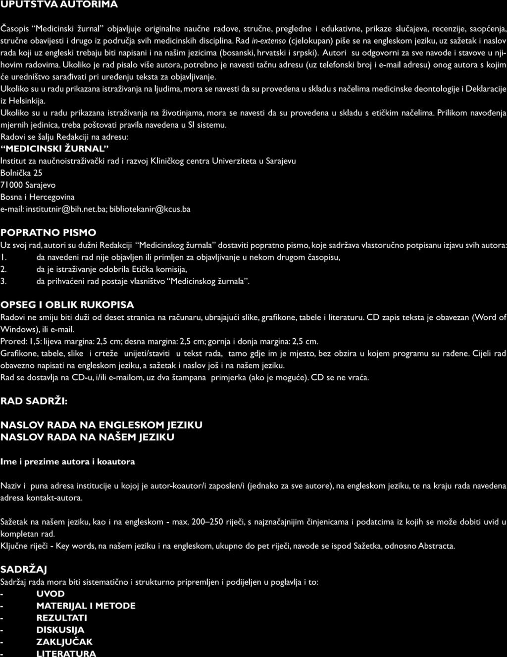
76

77 Uputstva autorima
78

79
ISSN СТАТИСТИКА СТАНОВНИШТВА ГОДИШЊЕ САОПШТЕЊЕ POPULATION STATISTICS ANNUAL RELEASE 25. IV Број/No. 115/19 РОЂЕНИ И УМРЛИ У РЕПУБ
 ISSN 2490-2950 2018 ГОДИШЊЕ САОПШТЕЊЕ POPULATION STATISTICS ANNUAL RELEASE 25. IV 2019. Број/No. 115/19 РОЂЕНИ И УМРЛИ У РЕПУБЛИЦИ СРПСКОЈ BIRTHS AND DEATHS IN REPUBLIKA SRPSKA Благи раст броја живорођених
ISSN СТАТИСТИКА СТАНОВНИШТВА ГОДИШЊЕ САОПШТЕЊЕ POPULATION STATISTICS ANNUAL RELEASE 25. IV Број/No. 121/18 РОЂЕНИ И УМРЛИ У РЕПУБ
 ISSN 2490-2950 2017 ГОДИШЊЕ САОПШТЕЊЕ POPULATION STATISTICS ANNUAL RELEASE 25. IV 2018. Број/No. 121/18 РОЂЕНИ И УМРЛИ У РЕПУБЛИЦИ СРПСКОЈ BIRTHS AND DEATHS IN REPUBLIKA SRPSKA Благи пад броја живорођених
ISSN СТАТИСТИКА СТАНОВНИШТВА ГОДИШЊЕ САОПШТЕЊЕ POPULATION STATISTICS ANNUAL RELEASE 25. IV Број/No. 107/17 РОЂЕНИ И УМРЛИ У РЕПУБ
 ISSN 2490-2950 2016 ГОДИШЊЕ САОПШТЕЊЕ POPULATION STATISTICS ANNUAL RELEASE 25. IV 2017. Број/No. 107/17 РОЂЕНИ И УМРЛИ У РЕПУБЛИЦИ СРПСКОЈ BIRTHS AND DEATHS IN REPUBLIKA SRPSKA Благи раст броја живорођених
F84 Zahtjev za priznavanje inostrane visokoškolske kvalifikacije (zaokružiti) Application for academic recognition of foreign higher qualification (ci
 Application for academic recognition of foreign higher qualification (ci") Zahtjev za priznavanje inostrane visokoškolske kvalifikacije (zaokružiti) Application for academic recognition of foreign higher qualification (circle) A. U SVRHU ZAPOŠLJAVANJA - STRUČNO PRIZNAVANJE /
Zahtjev za priznavanje inostrane visokoškolske kvalifikacije (zaokružiti) Application for academic recognition of foreign higher qualification (circle) A. U SVRHU ZAPOŠLJAVANJA - STRUČNO PRIZNAVANJE /
PowerPoint Presentation
 Како ревизије сврсисходности пословања утичу на рад корисника јавних средстава у Србији? Налази ревизија сврсисходности пословања као средства за унапређење вредности добијене за уложен новац Београд,
Adresa: Maršala Tita 9a/I Telefon: (033) Faks: (033) Web: Datum i vrijeme slanj
 Faks: (033) Web: Datum i vrijeme slanj") Adresa: Maršala Tita 9a/I Telefon: (033) 251-590 Faks: (033) 251-595 E-mail: ejn@javnenabavke.gov.ba Web: https://www.ejn.gov.ba Datum i vrijeme slanja obavještenja na objavu:27.6.2016. u 13:59 ISPRAVKA
Adresa: Maršala Tita 9a/I Telefon: (033) 251-590 Faks: (033) 251-595 E-mail: ejn@javnenabavke.gov.ba Web: https://www.ejn.gov.ba Datum i vrijeme slanja obavještenja na objavu:27.6.2016. u 13:59 ISPRAVKA
Adresa: Maršala Tita 9a/I Telefon: (033) Faks: (033) Web: Datum i vrijeme slanj
 Faks: (033) Web: Datum i vrijeme slanj") Adresa: Maršala Tita 9a/I Telefon: (033) 251-590 Faks: (033) 251-595 E-mail: ejn@javnenabavke.gov.ba Web: https://www.ejn.gov.ba Datum i vrijeme slanja obavještenja na objavu:27.6.2016. u 15:49 ISPRAVKA
Adresa: Maršala Tita 9a/I Telefon: (033) 251-590 Faks: (033) 251-595 E-mail: ejn@javnenabavke.gov.ba Web: https://www.ejn.gov.ba Datum i vrijeme slanja obavještenja na objavu:27.6.2016. u 15:49 ISPRAVKA
TITLE
 Travnik, 18.5.2017. Sveučilište Hercegovina Herzegovina University Institucionalni rani i predškolski odgoj i obrazovanje u BIH Institutional early and pre-school education in BIH Ograničenja obrazovnih
Acta medica Medianae (2000) 6 (21-25) PRETHODNA SAOPŠTENJA MORBIDITET OD AKUTNOG INFARKTA MIOKARDA U POPULACIJI ROMA GRADA NIŠA Marko LAZOVIĆ, Svetlan
 6 (21-25) PRETHODNA SAOPŠTENJA MORBIDITET OD AKUTNOG INFARKTA MIOKARDA U POPULACIJI ROMA GRADA NIŠA Marko LAZOVIĆ, Svetlan") Acta medica Medianae (2000) 6 (21-25) PRETHODNA SAOPŠTENJA MORBIDITET OD AKUTNOG INFARKTA MIOKARDA U POPULACIJI ROMA GRADA NIŠA i Milomir RANĐELOVIĆ Klinika za kardiovaskularne holesti Kliničkog centra
Acta medica Medianae (2000) 6 (21-25) PRETHODNA SAOPŠTENJA MORBIDITET OD AKUTNOG INFARKTA MIOKARDA U POPULACIJI ROMA GRADA NIŠA i Milomir RANĐELOVIĆ Klinika za kardiovaskularne holesti Kliničkog centra
HBIS GROUP SERBIA IRON & STEEL D.O.O. BEOGRAD CI Sektor Kontinualno poboljša je Continuous Improvement Sector..
 CI.. (Continuous Improvement CI) je eđu arod o priz ata metodologija implementirana 00. godi e u ašoj fa ri i Continuous Improvement (CI) is an internationally recognized methodology implemented in our
No type Здравствено, пензијско и инвалидско осигурање Health, pension and disability insurance 479
 No type 479 Методолошка објашњења Methodological explanations... 481 ТАБЕЛЕ TABLES.1. Здравствено осигурање Health insurance...483.2. Расходи здравственог осигурања Health insurance expenditures...483.3.
In accordance with Article 33 Paragraph 2 Item 4 of the Listing Rules of the Belgrade Stock Exchange j.s.c. Belgrade, NIS j.s.c. Novi Sad announces RE
 In accordance with Article 33 Paragraph 2 Item 4 of the Listing Rules of the Belgrade Stock Exchange j.s.c. Belgrade, NIS j.s.c. Novi Sad announces REPORT ON COMPANY BODY'S SESSION 126th NIS j.s.c. Board
No type Здравствено, пензијско и инвалидско осигурање Health, pension and disability insurance 507
 No type 507 Методолошка објашњења Methodological explanations... 509 ТАБЕЛЕ TABLES.1. Здравствено осигурање Health insurance...511.2. Расходи здравственог осигурања Health insurance expenditures...511.3.
CONFIDA_Mesecni_Newsletter_SRB_JUL
 BELGRADE ZAGREB LJUBLJANA VIENNA TIRANA SARAJEVO BANJA LUKA SKOPJE PODGORICA Your reference for Tax News in SERBIA July 2019. and international businesses with answers to key questions regarding tax regulations
ISSN СТАТИСТИКА ОБРАЗОВАЊА ГОДИШЊЕ САОПШТЕЊЕ ШКОЛСКА ГОДИНА/SCHOOL YEAR почетак/beginning of 2018/2019 EDUCATION STATISTICS ANNUAL RELEASE П
 ISSN 2490-2950 СТАТИСТИКА ОБРАЗОВАЊА ГОДИШЊЕ САОПШТЕЊЕ ШКОЛСКА ГОДИНА/SCHOOL YEAR почетак/beginning of 2018/2019 EDUCATION STATISTICS ANNUAL RELEASE Претходни подаци/preliminary data 1. IV 2019. Број/No.
Zdravstveno stanje stanovništva za godinu
 ЗДРАВСТВЕНО СТАЊE СТАНОВНИШТВА ЗА 2010. ГОДИНУ Analysis of health of population of Republic of Srpska 2010 Prilikom korištenja podataka iz ove publikacije obavezno navesti izvor, Institut za javno zdravstvo
Microsoft PowerPoint - Donacija-Zca [Compatibility Mode]
![Microsoft PowerPoint - Donacija-Zca [Compatibility Mode]](/thumbs/88/116330844.jpg "Microsoft PowerPoint - Donacija-Zca [Compatibility Mode]") Donacija naučnih časopisa i knjiga Zenica, 16.05.2013 Humanost u akciji (HIA) je međunarodna neprofitna obrazovna organizacija koja obrazuje, inspirira i i povezuje studente i mlade profesionalce u globalnu
Donacija naučnih časopisa i knjiga Zenica, 16.05.2013 Humanost u akciji (HIA) je međunarodna neprofitna obrazovna organizacija koja obrazuje, inspirira i i povezuje studente i mlade profesionalce u globalnu
MAZALICA DUŠKA.pdf
 SVEUČILIŠTE JOSIPA JURJA STROSSMAYERA U OSIJEKU ELEKTROTEHNIČKI FAKULTET Sveučilišni studij OPTIMIRANJE INTEGRACIJE MALIH ELEKTRANA U DISTRIBUCIJSKU MREŽU Diplomski rad Duška Mazalica Osijek, 2014. SADRŽAJ
No type Здравствено, пензијско и инвалидско осигурање Health, pension and disability insurance Статистички годишњак Републике Српске 2014 Statistical
 No type Статистички годишњак Републике Српске 2014 455 Методолошка објашњења Methodological explanations... 457 ТАБЕЛЕ TABLES.1. Здравствено осигурање Health insurance...459.2. Расходи здравственог осигурања
ZBORNIK APSTRAKTA BR. 1.ai
 I INTERNACIONALNI NAUČNI SIMPOZIJUM ULOGA I ZNAČAJ NOVE EKONOMIJE I INTERNATIONAL SCIENTIFIC SYMPOSIUM ROLE AND IMPORTANCE OF NEW ECONOMY BIJELJINA 01. OKTOBAR 2013 GODINE ORGANIZATOR: Oikos institut -
NN indd
 BROJ 81 STRANICA 9 PRILOG 1. Obrazac PISK-1 Zahtjev za priznavanje inozemnih stručnih kvalifikacija Application for recognition of professional qualifications Žensko Female Muško Male of profession 3.
Jasna Kellner
 1 Broji. 19 Ne pričaj. 37 Reci "Hvala". 2 Ne kasni. (Budi točan.) 20 Ne razumijem. 38 Reci "Molim". 3 Čitaj. 21 Ne spavaj. 39 Sjedni dolje. 4 Crtaj 22 Ne varaj. 40 Slušaj i ponovi. 5 Razumiješ li? 23 Obriši
In accordance with Article 33 Paragraph 2 Item 4 of the Listing Rules of the Belgrade Stock Exchange j.s.c. Belgrade, NIS j.s.c. Novi Sad announces RE
 In accordance with Article 33 Paragraph 2 Item 4 of the Listing Rules of the Belgrade Stock Exchange j.s.c. Belgrade, NIS j.s.c. Novi Sad announces REPORT ON COMPANY BODY'S SESSION 68th NIS j.s.c. Board
 4 CONTENTS 2 2 5 5 POD SOJOM U POVRŠINAMA NA RAZLIČITE VELIČINE U AP VOJVODINI 1 Bošnjak Danica, Rodić Vesna, Karapandžin Jelena 2 Rezime Veličina gazdinstva, iskazana raspoloživim poljoprivrednim zemljištem,
NN indd
 PETAK, 13. STUDENOGA 2015. NARODNE NOVINE BROJ 124 STRANICA 27 PRILOG 1. STRANICA 28 BROJ 124 NARODNE NOVINE PETAK, 13. STUDENOGA 2015. PRILOG 2. PETAK, 13. STUDENOGA 2015. NARODNE NOVINE BROJ 124 STRANICA
Dragi trkac i/ice, I ove godine imamo preporuku za vaš smještaj na Jahorini, ali ovog puta je to c ista eksluziva po STVARNO POSEBNIM cijenama u izuze
 Dragi trkac i/ice, I ove godine imamo preporuku za vaš smještaj na Jahorini, ali ovog puta je to c ista eksluziva po STVARNO POSEBNIM cijenama u izuzetnom hotelu Lavina s zvjezdice! Hotel ima izvrsnu hranu,
MODUL 12
 ANKETAO MJERENJU @IVOTNOG STANDARDA U BiH MODUL 11 Nepoljoprivredne aktivnosti KOMENTARI 1 NIVO FBiH 11.3.1 Procentualni raspored stanovni{tva koje posjeduje nepoljoprivredna preduze}a i polu vlasnika
Microsoft Word - CroDiab Registar 2009.doc
 HRVATSKI ZAVOD ZA JAVNO ZDRAVSTVO Zagreb, Rockefellerova 7. - SVEUČILIŠNA KLINIKA VUK VRHOVAC Dugi dol 4a, Zagreb IZVJEŠĆE ZA 2009. Nacionalni registar osoba sa šećernom bolešću CroDiab National Diabetes
VRAČEVIĆ FRANJO.pdf
 SVEUČILIŠTE JOSIPA JURJA STROSSMAYERA U OSIJEKU ELEKTROTEHNIČKI FAKULTET Sveučilišni studij IZRADA PROJEKTA AKTIVNE ENERGETSKI NEOVISNE KUĆE Diplomski rad Franjo Vračević Osijek, 2015 godina. SADRŽAJ 1.
ISSN СТАТИСТИКА ОБРАЗОВАЊА ГОДИШЊЕ САОПШТЕЊЕ EDUCATION STATISTICS ANNUAL RELEASE 1. IV Број/No. 88/19 ОБРАЗОВАЊЕ У САОБРАЋАЈУ EDU
 ISSN 2490-2950 2018 СТАТИСТИКА ОБРАЗОВАЊА ГОДИШЊЕ САОПШТЕЊЕ EDUCATION STATISTICS ANNUAL RELEASE 1. IV 2019. Број/No. 88/19 ОБРАЗОВАЊЕ У САОБРАЋАЈУ EDUCATION IN TRAFFIC У 2018. години, 217 ауто-школa обављало
In accordance with Article 33 Paragraph 2 Item 4 of the Listing Rules of the Belgrade Stock Exchange j.s.c. Belgrade, NIS j.s.c. Novi Sad announces RE
 In accordance with Article 33 Paragraph 2 Item 4 of the Listing Rules of the Belgrade Stock Exchange j.s.c. Belgrade, NIS j.s.c. Novi Sad announces REPORT ON COMPANY BODY'S SESSION LXXIX NIS j.s.c. Board
Microsoft Word - CroDiab Registar 2008.doc
 HRVATSKI ZAVOD ZA JAVNO ZDRAVSTVO Zagreb, Rockefellerova 7. - SVEUČILIŠNA KLINIKA VUK VRHOVAC Dugi dol 4a, Zagreb IZVJEŠĆE ZA 2008. Nacionalni registar osoba sa šećernom bolešću CroDiab National Diabetes
ISSN /2018 СТАТИСТИКА ОБРАЗОВАЊА EDUCATION STATISTICS ГОДИШЊЕ САОПШТЕЊЕ ANNUAL RELEASE Претходни подаци/preliminary data 10. III Б
 ISSN 2490-2950 2017/2018 СТАТИСТИКА ОБРАЗОВАЊА EDUCATION STATISTICS ГОДИШЊЕ САОПШТЕЊЕ ANNUAL RELEASE Претходни подаци/preliminary data 10. III 2018. /No. 57/18 ПРЕДШКОЛСКЕ УСТАНОВЕ У ШКОЛСКОЈ 2017/2018.
In accordance with Article 33 Paragraph 2 Item 4 of the Listing Rules of the Belgrade Stock Exchange j.s.c. Belgrade, NIS j.s.c. Novi Sad announces RE
 In accordance with Article 33 Paragraph 2 Item 4 of the Listing Rules of the Belgrade Stock Exchange j.s.c. Belgrade, NIS j.s.c. Novi Sad announces REPORT ON COMPANY BODY'S SESSION 63rd NIS j.s.c. Board
ISSN /2019 СТАТИСТИКА ОБРАЗОВАЊА EDUCATION STATISTICS ГОДИШЊЕ САОПШТЕЊЕ ANNUAL RELEASE Претходни подаци/preliminary data 11. III Б
 ISSN 2490-2950 2018/2019 СТАТИСТИКА ОБРАЗОВАЊА EDUCATION STATISTICS ГОДИШЊЕ САОПШТЕЊЕ ANNUAL RELEASE Претходни подаци/preliminary data 11. III 2019. /No. 61/19 ПРЕДШКОЛСКЕ УСТАНОВЕ У ШКОЛСКОЈ 2018/2019.
Odabrana poglavlja iz programskog inženjerstva
 OPIPI odabrana poglavlja iz programskog inženjerstva distributed software developmentdsd Mario Žagar Kolegij OPIPI/DSD Sveučilište u Zagrebu Fakultet elektrotehnike i računarstva Mälardalen University
46th Croatian & 6th International Symposium on Agriculture
 IZVORNI ZNANSTVENI RAD Smanjenje prinosa poljoprivrednih kultura u uvjetima bez navodnjavanja na području sjeveroistočne Bosne Univerzitet u Sarajevu, Poljoprivredno-prehrambeni fakultet, Zmaja od Bosne
SG/NJ 2018;23:13-7 DOI: /sgnj Mišljenje medicinskih sestara i tehničara o metodama vođenja sestrinske dokumentacije-iskustva žu
 DOI: 10.11608/sgnj.2018.23.004 13 Mišljenje medicinskih sestara i tehničara o metodama vođenja sestrinske dokumentacije-iskustva županijske bolnice Opinion of nurses and technicians about the methods of
No Slide Title
 Akreditacija laboratorijskih pretraga - sadašnjost ili budućnost? Dr. sc. Sonja Perkov, spec.medicinske biokemije Klinički zavod za medicinsku biokemiju i laboratorijsku medicinu, Kliničke bolnice Merkur
Slide 1
 Strateški pristup u razvoju poslovanja Dražen Oreščanin Poslovna inteligencija d.o.o. Predsjednik Uprave Poslovna inteligencija je... HRVATSKA - 2001 SRBIJA- 2006 CRNA GORA - 2008 SLOVENIJA - 2010 BIH
In accordance with Article 33 Paragraph 2 Item 4 of the Listing Rules of the Belgrade Stock Exchange j.s.c. Belgrade, NIS j.s.c. Novi Sad announces RE
 In accordance with Article 33 Paragraph 2 Item 4 of the Listing Rules of the Belgrade Stock Exchange j.s.c. Belgrade, NIS j.s.c. Novi Sad announces REPORT ON COMPANY BODY'S SESSION 132nd NIS j.s.c. Board
KATUŠIĆ ANTONIO.pdf
 SVEUILIŠTE JOSIP JURAJ STROSSMAYER ELEKTROTEHNIKI FAKULTET OSIJEK Preddiplomski studij raunarstva PROGRAMSKI JEZIK RUBY ZAVRŠNI RAD Antonio Katuši OSIJEK, svibanj 2015. SVEUILIŠTE JOSIP JURAJ STROSSMAYER
НИС a.д. Нови Сад / NIS j.s.c. Novi Sad Народног фронта 12 / Narodnog fronta 12 СКУПШТИНА АКЦИОНАРА / SHAREHOLDERS ASSEMBLY Број (No.): 70/SA-od/VIII-
: 70/SA-od/VIII-") НИС a.д. Нови Сад / NIS j.s.c. Novi Sad Народног фронта 12 / Narodnog fronta 12 СКУПШТИНА АКЦИОНАРА / SHAREHOLDERS ASSEMBLY Број (No.): 70/SA-od/VIII-2/5 Датум (Date): 28.06.2016 На основу члана 329. став
НИС a.д. Нови Сад / NIS j.s.c. Novi Sad Народног фронта 12 / Narodnog fronta 12 СКУПШТИНА АКЦИОНАРА / SHAREHOLDERS ASSEMBLY Број (No.): 70/SA-od/VIII-2/5 Датум (Date): 28.06.2016 На основу члана 329. став
Safety with Car Seats and Booster Seats - Bosnian
 Safety with Car Seats and Booster Seats Car crashes are the main cause of accidental death and serious injury of children. Correctly using a car or booster seat can save your child s life. Throughout the
PowerPoint Presentation
 Žan Mone program Eras Nacionalni Erasmus + info dan, 23. novembar 2017. Univerzitet Crne Gore Šta je Žan Mone program Program Žan Mone (1989) - uvođenje evropskih studija Posvećeni uspomeni na Žan Monea
Adresa: Maršala Tita 9a/I Telefon: (033) Faks: (033) Web: Datum i vrijeme slanj
 Faks: (033) Web: Datum i vrijeme slanj") Adresa: Maršala Tita 9a/I Telefon: (033) 251-590 Faks: (033) 251-595 E-mail: ejn@javnenabavke.gov.ba Web: https://www.ejn.gov.ba Datum i vrijeme slanja obavještenja na objavu:7.2.2019. u 10:21 OBAVJEŠTENJE
Adresa: Maršala Tita 9a/I Telefon: (033) 251-590 Faks: (033) 251-595 E-mail: ejn@javnenabavke.gov.ba Web: https://www.ejn.gov.ba Datum i vrijeme slanja obavještenja na objavu:7.2.2019. u 10:21 OBAVJEŠTENJE
ISSN СТАТИСТИКА ОБРАЗОВАЊА ГОДИШЊЕ САОПШТЕЊЕ Ш К О Л С К А Г О Д И Н А / S C H O O L Y E A R почетак/beginning of 2017/2018 EDUCATION STATIS
 ISSN 2490-2950 СТАТИСТИКА ОБРАЗОВАЊА ГОДИШЊЕ САОПШТЕЊЕ Ш К О Л С К А Г О Д И Н А / S C H O O L Y E A R почетак/beginning of 2017/2018 EDUCATION STATISTICS ANNUAL RELEASE Претходни подаци/preliminary data
InfoXgen Input Evaluation Königsbrunnerstr Enzersfeld Austria Austria Bio Garantie d.o.o. Rudera Boškovica Cako
 Bio Garantie d.o.o. Confirmation Confirmation of conformity with Council Regulation (EC) No 834/2007 on organic production and labelling of organic products as amended. After evaluation of the documents
Adresa: Maršala Tita 9a/I Telefon: (033) Faks: (033) Web: Datum i vrijeme slanj
 Faks: (033) Web: Datum i vrijeme slanj") Adresa: Maršala Tita 9a/I Telefon: (033) 251-590 Faks: (033) 251-595 E-mail: ejn@javnenabavke.gov.ba Web: https://www.ejn.gov.ba Datum i vrijeme slanja obavještenja na objavu:19.12.2017. u 14:31 OBAVJEŠTENJE
Adresa: Maršala Tita 9a/I Telefon: (033) 251-590 Faks: (033) 251-595 E-mail: ejn@javnenabavke.gov.ba Web: https://www.ejn.gov.ba Datum i vrijeme slanja obavještenja na objavu:19.12.2017. u 14:31 OBAVJEŠTENJE
No type Социјална заштита Social welfare Статистички годишњак Републике Српске 2014 Statistical Yearbook of Republika Srpska
 No type Статистички годишњак Републике Српске 2014 Statistical Yearbook of Republika Srpska 2014 483 Методолошка објашњења Methodological explanations... 485 ТАБЕЛЕ TABLES.1. Корисници, облици и услуге
29 Правосуђе Administration of justice Методолошка објашњења Methodological explanations Извори и методe прикупљања података Подаци о пунољетним и мал
 Методолошка објашњења Methodological explanations Извори и методe прикупљања података Подаци о пунољетним и малољетним учиниоцома кривичних дјела добијени су статистичким извјештајима које подносе тужилаштва,
No type Социјална заштита Social welfare 515
 No type 515 Методолошка објашњења Methodological explanations... 517 ТАБЕЛЕ TABLES.1. Корисници, облици и услуге социјалне заштите Beneficiaries, forms and services of social welfare...519.2. Малољетни
AM_Ple_LegReport
 6.12.2018 A8-0417/158 Amandman 158 Uvodna izjava 21. (21) Studije, uključujući ispitivanja, koje poslovni subjekti podnose u sklopu zahtjeva za odobrenje na temelju sektorskih propisa Unije o hrani obično
Microsoft Word - reprint prelom1.doc
 Pretisci iz graditeljskog tiska VIESTI KLUBA INŽINIRA I ARHITEKTA IV. (1883), 1, 1-2 Prof. dr. sc. Dražen Aničić, dipl. ing. građ. redoviti član Akademije tehničkih znanosti Hrvatske SUSTAV JAVNIH STUBA
Microsoft Word MET.DOC
 8. METODOLOŠKA OBJAŠNJENJA Izvori i metode prikupljanja podataka Podaci o nezaposlenosti i zapošljavanju preuzeti su od Hrvatskog zavoda za zapošljavanje. Podaci o nezaposlenosti i zapošljavanju temelje
INTERNATIONAL UNIVERSITY OF SARAJEVO INTERNACIONALNI UNIVERZITET U SARAJEVU - SENAT - Number: IUS-SENAT /14 Sarajevo, April 4, 2014 Pursuant
 - SENAT - Number: IUS-SENAT 11-1044-2/14 Sarajevo, April 4, 2014 Pursuant to Article 53, Paragraph (3) and Article 108, Paragraph (9) of the Law on Higher Education ( Official Gazette of Canton Sarajevo,
No type Правосуђе Administration of justice Статистички годишњак Републике Српске 2014 Statistical Yearbook of Republika Srpska
 No type Статистички годишњак Републике Српске 2014 473 Методолошка објашњења Methodological explanations... 475 ТАБЕЛЕ TABLES.1. Судови опште надлежности Courts of general jurisdiction...477.2. Судови
januar siječanj JANUARY Greške se mogu ispraviti. Even if you make a mistake, you can fix it.
 januar siječanj JANUARY 2020. Greške se mogu ispraviti. Even if you make a mistake, you can fix it. Naslov: Probudi, pokreni, promijeni! Izdavač: TPO Fondacija Ilustracije i dizajn: Neven Misaljević Štampa:
PRILOG I. PONUDBENI LIST S DODACIMA ZA ZAJEDNICU PONUDITELJA I PODIZVODITELJE / APPENDIX I. BIDDING LIST WITH APPENDICES FOR JOINT BIDDERS AND SUB- CO
 PRILOG I. PONUDBENI LIST S DODACIMA ZA ZAJEDNICU PONUDITELJA I PODIZVODITELJE / APPENDIX I. BIDDING LIST WITH APPENDICES FOR JOINT BIDDERS AND SUB- CONTRACTORS Predmet nabave / Subject of procurement:
Pisanje predloga projekta
 MINISTARSTVO ZA NAUKU I TEHNOLOŠKI RAZVOJ Konsultativni biro za meñunarodne projekte Sedmi okvirni program EU za istraživanje i razvoj Kako učestvovati u projektima O čemu će biti reči... Učešće MSP u
Slide 1
 Activity of creation of courses for professional insertion, career guidance and internship Natali Stojiljkovic (AQ)/Tatjana Anđelković (UN) ICT Networking for Overcoming Technical and Social Barriers in
Microsoft Word - akademsko priznavanje-konacno.doc
 SVEUČILIŠTE U ZADRU / UNIVERSITY OF ZADAR Ured za akademsko priznavanje inozemnih visokoškolskih kvalifikacija / Academic Recognition Office Mihovila Pavlinovića bb HR 23 000 ZADAR Zahtjev za priznavanje
Master slide prezentacija
 Javnozdravstveni problem premalignih i malignih lezija vrata maternice u Republici Hrvatskoj Ante Ćorušić Zagreb,23. svibnja 2018. Cervical Cancer Second Most Frequent Cancer in Young European Women 1
No type Буџети и фондови Budgets and funds 155
 No type 155 Методолошка објашњења Methodological explanations... 157 ТАБЕЛЕ TABLES.1. Остварени приходи и расходи буџета Републике Realised budgetary revenues and expenditures of Republika Srpska...158.2.
n50
 N50LUSK Vodič za ažuriranje TV softvera TV Software Update Guide Hrvatski vodič za ažuriranje 1. Korak Provjerite ime modela na stražnjoj strani TV-a. Prije preuzimanja softvera za ažuriranje, molimo provjerite
SIGURNOSNI NALOG BROJ: 2017/001 rev 01 SAFETY ORDER NUMBER: 2017/001 rev 01 Naslov: Sigurnosni nalog kojim se utvrđuju uslovi za upotrebu sistema besp
 SIGURNOSNI NALOG BROJ: 2017/001 rev 01 SAFETY ORDER NUMBER: 2017/001 rev 01 Naslov: Sigurnosni nalog kojim se utvrđuju uslovi za upotrebu sistema bespilotnih vazduhoplova i vazduhoplovnih modela u oblastima
ВЕЋУ МЕНТОРА
 НАСТАВНО-НАУЧНОМ ВЕЋУ ФАКУЛТЕТА МЕДИЦИНСКИХ НАУКА УНИВЕРЗИТЕТА У КРАГУЈЕВЦУ Комисија за припрему извештаја у саставу: 1. проф. др Сања Коцић, ванредни професор за ужу научну област Социјална медицина Факултета